
solo para uso en investigación
GNF-2 Bcr-Abl inhibidor
Cat. No.S2899
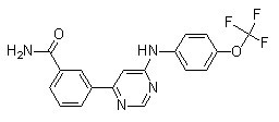
Estructura química
Peso molecular: 374.32
Control de calidad
| Dianas relacionadas | EGFR VEGFR JAK PDGFR FGFR Src HIF FLT FLT3 HER2 |
|---|---|
| Otros Bcr-Abl Inhibidores | Degrasyn (WP1130) Bafetinib Rebastinib (DCC-2036) GNF-5 Berbamine Olverembatinib (GZD824) dimesylate PD173955 GNF-7 Radotinib Berbamine dihydrochloride |
Información química, almacenamiento y estabilidad
| Peso molecular | 374.32 | Fórmula | C18H13F3N4O2 |
Almacenamiento (Desde la fecha de recepción) | |
|---|---|---|---|---|---|
| Nº CAS | 778270-11-4 | Descargar SDF | Almacenamiento de soluciones madre |
|
|
| Sinónimos | N/A | Smiles | C1=CC(=CC(=C1)C(=O)N)C2=CC(=NC=N2)NC3=CC=C(C=C3)OC(F)(F)F | ||
Solubilidad
|
In vitro |
DMSO
: 74 mg/mL
(197.69 mM)
Water : Insoluble Ethanol : Insoluble |
Calculadora de Molaridad
|
In vivo |
|||||
Calculadora de formulación in vivo (Solución clara)
Paso 1: Introduzca la información a continuación (Recomendado: Un animal adicional para tener en cuenta la pérdida durante el experimento)
Paso 2: Introduzca la formulación in vivo (Esto es solo la calculadora, no la formulación. Por favor, contáctenos primero si no hay una formulación in vivo en la sección de Solubilidad.)
Resultados del cálculo:
Concentración de trabajo: mg/ml;
Método para preparar el líquido maestro de DMSO: mg fármaco predissuelto en μL DMSO ( Concentración del líquido maestro mg/mL, Por favor, contáctenos primero si la concentración excede la solubilidad del DMSO del lote del fármaco. )
Método para preparar la formulación in vivo: Tomar μL DMSO líquido maestro, luego añadirμL PEG300, mezclar y clarificar, luego añadirμL Tween 80, mezclar y clarificar, luego añadir μL ddH2O, mezclar y clarificar.
Método para preparar la formulación in vivo: Tomar μL DMSO líquido maestro, luego añadir μL Aceite de maíz, mezclar y clarificar.
Nota: 1. Por favor, asegúrese de que el líquido esté claro antes de añadir el siguiente disolvente.
2. Asegúrese de añadir el (los) disolvente(s) en orden. Debe asegurarse de que la solución obtenida, en la adición anterior, sea una solución clara antes de proceder a añadir el siguiente disolvente. Se pueden utilizar métodos físicos como el vórtice, el ultrasonido o el baño de agua caliente para ayudar a la disolución.
Mecanismo de acción
| Targets/IC50/Ki |
Bcr-Abl (SUP-B15 cell line)
268 nM
Bcr-Abl (K562 cell line)
273 nM
|
|---|---|
| In vitro |
GNF-2 provoca una inhibición del crecimiento dependiente de la dosis en las líneas celulares Bcr-abl-positivas con valores de IC50 de 273 nM (K562) y 268 nM (SUP-B15). Este compuesto inhibe el crecimiento de las células Ba/F3.p210E255V y Ba/F3.p185Y253H con valores de IC50 de 268 nM y 194 nM respectivamente. Este químico (1 μM) induce la apoptosis de las células Ba/F3.p210, así como de las células Ba/F3.p210E255V. Inhibe la fosforilación de tirosina celular de Bcr-abl de manera dependiente de la dosis con una IC50 de 267 nM. Este compuesto (1 μM) induce una disminución significativa en los niveles de fosfo-Stat5 en las células Ba/F3.p210. Se une al bolsillo de unión mirística de Bcr-abl. Este inhibidor inhibe la actividad quinasa de c-Abl no miristilado más potentemente que la de c-Abl miristilado al unirse al bolsillo de unión a miristato en el lóbulo C del dominio quinasa. Este agente (10 μM) requiere BCR y/o los dominios SH3 y/o SH2 de c-Abl para inhibir la proliferación celular dependiente de BCR-Abl. Este, pero no un análogo GNF-2 metilado, se une a c-Abl en extractos celulares derivados de fibroblastos 3T3. Este compuesto (10 μM), de manera dependiente de la dosis, inhibe claramente la fosforilación de tirosina de CrkII. Inhibe la fosforilación de CrkII en células que expresan c-AblG2A con una IC50 de 0.051 μM. Este químico se une en una conformación extendida en el bolsillo de miristato con el grupo CF3 enterrado a la misma profundidad que los dos carbonos finales del ligando de miristato. Este compuesto (10 µM) combinado con imatinib (1 µM) reduce el número de clones resistentes a 1 µM de imatinib en al menos un 90 %. Inhibe la autofosforilación y proliferación de células BafF3 que expresan mutantes p210Bcr-Abl y p210Bcr-Abl. Este agente (8 nM) en combinación con GNF-5 (20 nM) produce efectos aditivos con respecto a la inhibición de la actividad quinasa de Abl64-515.
|
| Ensayo de quinasa |
Ensayo de unión
|
|
Las proteínas recombinantes (100 nM para cada construcción) o las proteínas inmunoprecipitadas se diluyen en tampón de quinasa (20 mM HEPES (pH 7.4), 50 mM KCl, 0.1% CHAPS, 30 mM MgCl2, 2 mM MnCl2, 1 mM DTT y 1% de glicerol). Alícuotas de las proteínas diluidas se preincuban con DMSO o este compuesto durante 30 min a temperatura ambiente y luego se añaden a las placas de reacción K-LISA PTK EAY. La reacción de la quinasa se inicia añadiendo 0.1 mM de ATP y se deja proceder durante 30 min a temperatura ambiente. La fosforilación de GST-Abltide se monitoriza mediante SDS-PAGE y análisis de fosforimágenes o autorradiografía.
|
Referencias |
|
Soporte técnico
Tel: +1-832-582-8158 Ext:3
Si tiene alguna otra consulta, por favor deje un mensaje.

Los productos son solo para uso de investigación. No para uso humano. No vendemos a pacientes.
©Copyright 2013 Selleck Chemicals. Todos los derechos reservados.