
solo para uso en investigación
NVP-AEW541 IGF-1R inhibidor
Cat. No.S1034
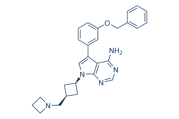
Estructura química
Peso molecular: 439.55
Control de calidad
| Dianas relacionadas | EGFR VEGFR PDGFR FGFR c-Met Src MEK CSF-1R FLT3 HER2 |
|---|---|
| Otros IGF-1R Inhibidores | Linsitinib (OSI-906) BMS-536924 BMS-754807 Picropodophyllin (AXL1717) GSK1904529A AG-1024 NVP-ADW742 NT157 PQ 401 MSDC-0160 |
Cultivo celular, tratamiento y concentración de trabajo
| Líneas celulares | Tipo de ensayo | Concentración | Tiempo de incubación | Formulación | Descripción de la actividad | PMID |
|---|---|---|---|---|---|---|
| NWT-21 | Kinase assay | ~10 μM | DMSO | inhibits IGF-IR with IC50 of 0.086 ± 0.028 μM | 15050915 | |
| A14 | Kinase assay | ~10 μM | DMSO | inhibits InsR with IC50 of 2.3 ± 0.163 μM | 15050915 | |
| A431 | Kinase assay | ~10 μM | DMSO | inhibits HER1 with IC50 of >10 μM | 15050915 | |
| A31 | Kinase assay | ~10 μM | DMSO | inhibits PDGFR with IC50 of >10 μM | 15050915 | |
| GIST882 | Kinase assay | ~10 μM | DMSO | inhibits c-Kit with IC50 of >5 μM | 15050915 | |
| 32D-Bcr-Abl | Kinase assay | ~10 μM | DMSO | inhibits Bcr-Abl p210 with IC50 of >10 μM | 15050915 | |
| MCF-7 | Cytoxicity assay | DMSO | IC50=1.64 μM | 15050915 | ||
| NWT-21 | Growth inhibitory assay | DMSO | IC50=0.163 μM | 15050915 | ||
| TC-71 | Growth inhibitory assay | ~1 μM | DMSO | inhibits insulin-like growth factor-I–mediated growth | 15867386 | |
| TC-71 | Growth inhibitory assay | ~7 μM | DMSO | IC50<0.5 μM | 15867386 | |
| Saos-2 | Growth inhibitory assay | ~7 μM | DMSO | IC50<3 μM | 15867386 | |
| U-2OS | Growth inhibitory assay | ~7 μM | DMSO | IC50<0.5 μM | 15867386 | |
| SK-ES-1 | Growth inhibitory assay | ~7 μM | DMSO | IC50<0.5 μM | 15867386 | |
| SK-N-MC | Growth inhibitory assay | ~7 μM | DMSO | IC50<0.5 μM | 15867386 | |
| RD-ES | Growth inhibitory assay | ~7 μM | DMSO | IC50<0.5 μM | 15867386 | |
| SJ-Rh 30 | Growth inhibitory assay | ~7 μM | DMSO | IC50<0.5 μM | 15867386 | |
| SJ-Rh 4 | Growth inhibitory assay | ~7 μM | DMSO | IC50<0.5 μM | 15867386 | |
| 6647 | Growth inhibitory assay | ~7 μM | DMSO | IC50<0.5 μM | 15867386 | |
| SARG | Growth inhibitory assay | ~7 μM | DMSO | IC50<3 μM | 15867386 | |
| MOS | Growth inhibitory assay | ~7 μM | DMSO | IC50<4 μM | 15867386 | |
| IOR/OS7 | Growth inhibitory assay | ~7 μM | DMSO | IC50<1 μM | 15867386 | |
| IOR/OS9 | Growth inhibitory assay | ~7 μM | DMSO | IC50<6 μM | 15867386 | |
| IOR/OS10 | Growth inhibitory assay | ~7 μM | DMSO | IC50<5 μM | 15867386 | |
| IOR/OS14 | Growth inhibitory assay | ~7 μM | DMSO | IC50<4 μM | 15867386 | |
| LAP35 | Growth inhibitory assay | ~7 μM | DMSO | IC50<0.5 μM | 15867386 | |
| IOR/BRZ | Growth inhibitory assay | ~7 μM | DMSO | IC50<0.5 μM | 15867386 | |
| IOR/CAR | Growth inhibitory assay | ~7 μM | DMSO | IC50<1 μM | 15867386 | |
| IOR/NGR | Growth inhibitory assay | ~7 μM | DMSO | IC50<0.5 μM | 15867386 | |
| IOR/RCH | Growth inhibitory assay | ~7 μM | DMSO | IC50<0.5 μM | 15867386 | |
| RMZ-RC2 | Growth inhibitory assay | ~7 μM | DMSO | IC50<0.5 μM | 15867386 | |
| CCA | Growth inhibitory assay | ~7 μM | DMSO | IC50<2 μM | 15867386 | |
| RD/18 | Growth inhibitory assay | ~7 μM | DMSO | IC50<4 μM | 15867386 | |
| OVCAR-3 | Growth inhibitory assay | ~15 μM | DMSO | inhibits cell proliferation | 16300820 | |
| OVCAR-4 | Growth inhibitory assay | ~15 μM | DMSO | inhibits cell proliferation | 16300820 | |
| OVCAR-3 | Apoptosis assay | ~15 μM | DMSO | induces apoptosis | 16300820 | |
| OVCAR-4 | Apoptosis assay | ~15 μM | DMSO | induces apoptosis | 16300820 | |
| OVCAR-3 | Function assay | ~15 μM | DMSO | Decreases phosphorylation of AKT | 16300820 | |
| Huh-7 | Growth inhibitory assay | ~10 μM | DMSO | IC50=1.4 μM | 16530734 | |
| Hep-G2 | Growth inhibitory assay | ~10 μM | DMSO | IC50=1.8 μM | 16530734 | |
| Hep-3B | Growth inhibitory assay | ~10 μM | DMSO | IC50=1.9 μM | 16530734 | |
| SK-Hep-1 | Growth inhibitory assay | ~10 μM | DMSO | IC50=6.9 μM | 16530734 | |
| Huh-7 | Function assay | ~10 μM | DMSO | Induces cell cycle arrest | 16530734 | |
| Hep-G2 | Function assay | ~10 μM | DMSO | Induces cell cycle arrest | 16530734 | |
| SK-Hep-1 | Function assay | ~10 μM | DMSO | Induces cell cycle arrest | 16530734 | |
| BON | Kinase assay | ~6 μM | DMSO | induces dephosphorylation of IGF-1R | 16601284 | |
| BON | Growth inhibitory assay | ~10 μM | DMSO | IC50=6.6 μM | 16601284 | |
| CM | Growth inhibitory assay | ~5 μM | DMSO | IC50=3.3 μM | 16601284 | |
| BON | Function assay | ~7.5 μM | DMSO | induces cell cycle arrest | 16601284 | |
| CM | Function assay | ~5 μM | DMSO | induces cell cycle arrest | 16601284 | |
| BON | Apoptosis assay | ~7.5 μM | DMSO | induces Apoptosis | 16601284 | |
| CM | Apoptosis assay | ~5 μM | DMSO | induces Apoptosis | 16601284 | |
| HT-29 | Growth inhibitory assay | ~10 μM | DMSO | IC50=1.7 μM | 17007015 | |
| HCT-116 | Growth inhibitory assay | ~10 μM | DMSO | IC50=2.5 μM | 17007015 | |
| primary colorectal cancer cells | Function assay | ~5 μM | DMSO | alters the morphology of the remaining cells | 17007015 | |
| HTLA-230 | Function assay | ~8 μM | DMSO | inhibits IGF-II-mediated stimulation of IGF-IR and Akt | 17121898 | |
| KCNR | Function assay | ~8 μM | DMSO | inhibits IGF-II-mediated stimulation of IGF-IR and Akt | 17121898 | |
| SK-N-BE2c | Function assay | ~8 μM | DMSO | inhibits IGF-II-mediated stimulation of IGF-IR and Akt | 17121898 | |
| SK-N-BE | Function assay | ~8 μM | DMSO | inhibits IGF-II-mediated stimulation of IGF-IR and Akt | 17121898 | |
| LAN-5 | Function assay | ~8 μM | DMSO | inhibits IGF-II-mediated stimulation of IGF-IR and Akt | 17121898 | |
| GI-CA-N | Function assay | ~8 μM | DMSO | inhibits IGF-II-mediated stimulation of IGF-IR and Akt | 17121898 | |
| SH-EP | Function assay | ~8 μM | DMSO | inhibits IGF-II-mediated stimulation of IGF-IR and Akt | 17121898 | |
| SK-N-AS | Function assay | ~8 μM | DMSO | inhibits IGF-II-mediated stimulation of IGF-IR and Akt | 17121898 | |
| RN-GA | Function assay | ~8 μM | DMSO | inhibits IGF-II-mediated stimulation of IGF-IR and Akt | 17121898 | |
| SY-5Y(N) | Function assay | ~8 μM | DMSO | inhibits IGF-II-mediated stimulation of IGF-IR and Akt | 17121898 | |
| GI-CA-N | Growth inhibitory assay | ~8 μM | DMSO | IC50= 6.8 μM | 17121898 | |
| SH-EP | Growth inhibitory assay | ~8 μM | DMSO | IC50= 3 μM | 17121898 | |
| HTLA-230 | Growth inhibitory assay | ~8 μM | DMSO | IC50= 0.5 μM | 17121898 | |
| SK-N-BE2c | Growth inhibitory assay | ~8 μM | DMSO | IC50= 1.1 μM | 17121898 | |
| SK-N-BE2 | Growth inhibitory assay | ~8 μM | DMSO | IC50= 3 μM | 17121898 | |
| SY-5Y (N) | Growth inhibitory assay | ~8 μM | DMSO | IC50= 2.4 μM | 17121898 | |
| LAN-5 | Growth inhibitory assay | ~8 μM | DMSO | IC50= 0.4 μM | 17121898 | |
| KCNR | Growth inhibitory assay | ~8 μM | DMSO | IC50= 0.4 μM | 17121898 | |
| RN-GA | Growth inhibitory assay | ~8 μM | DMSO | IC50= 1.3 μM | 17121898 | |
| SK-N-AS | Growth inhibitory assay | ~8 μM | DMSO | induces apoptosis | 17121898 | |
| KCNR | Apoptosis assay | ~8 μM | DMSO | induces apoptosis | 17121898 | |
| GI-CA-N | Apoptosis assay | ~8 μM | DMSO | induces apoptosis | 17121898 | |
| HTLA-230 | Apoptosis assay | ~8 μM | DMSO | induces apoptosis | 17121898 | |
| SK-N-BE2c | Apoptosis assay | ~8 μM | DMSO | induces apoptosis | 17121898 | |
| SY-5Y (N) | Apoptosis assay | ~8 μM | DMSO | induces apoptosis | 17121898 | |
| HL60AR | Function assay | 160 nM | enhances the levels of p27Kip1 | 17361225 | ||
| HL60AR | Apoptosis assay | ~200 nM | induces apoptosis | 17361225 | ||
| HPAF-II | Kinase assay | ~1 μM | DMSO | inhibits IGF-I-mediated signalling | 18445520 | |
| HPAF-II | Growth inhibitory assay | ~2 μM | DMSO | inhibits cell proliferation | 18445520 | |
| HPAF-II | Function assay | ~2 μM | DMSO | inhibits basal and IGF-I-mediated pancreatic cancer cell migration | 18445520 | |
| TFK-1 | Growth inhibitory assay | ~250 nM | DMSO | IC50=0.26 μM | 20066734 | |
| EGI-1 | Growth inhibitory assay | ~250 nM | DMSO | IC50=0.28 μM | 20066734 | |
| CC-LP-1 | Growth inhibitory assay | ~250 nM | DMSO | IC50=0.15 μM | 20066734 | |
| CC-SW-1 | Growth inhibitory assay | ~250 nM | DMSO | IC50=0.54 μM | 20066734 | |
| Sk-ChA-1 | Growth inhibitory assay | ~250 nM | DMSO | IC50=0.2 μM | 20066734 | |
| Mz-ChA-1 | Growth inhibitory assay | ~250 nM | DMSO | IC50=1.39 μM | 20066734 | |
| Mz-ChA-2 | Growth inhibitory assay | ~250 nM | DMSO | IC50=0.73 μM | 20066734 | |
| ECC-1 | Kinase assay | ~10 μM | DMSO | inhibits IGF-IR activation by 98% | 21295335 | |
| Ishikawa | Kinase assay | ~10 μM | DMSO | inhibits IGF-IR activation by 93% | 21295335 | |
| USPC-1 | Kinase assay | ~10 μM | DMSO | inhibits IGF-IR activation by 100% | 21295335 | |
| USPC-2 | Kinase assay | ~10 μM | DMSO | inhibits IGF-IR activation by 96% | 21295335 | |
| ECC-1 | Growth inhibitory assay | ~10 μM | DMSO | decreases cell proliferation | 21295335 | |
| Ishikawa | Growth inhibitory assay | ~10 μM | DMSO | decreases cell proliferation | 21295335 | |
| USPC-1 | Growth inhibitory assay | ~10 μM | DMSO | decreases cell proliferation | 21295335 | |
| USPC-2 | Growth inhibitory assay | ~10 μM | DMSO | decreases cell proliferation | 21295335 | |
| Ba/F3 | Function assay | Inhibition of full length IGF-1 receptor (unknown origin) transfected in Ba/F3 cells assessed as cell proliferation, IC50=0.02μM | 26951753 | |||
| HEK293 | Function assay | 60 mins | Inhibition of full length IGF-1 receptor (unknown origin) autophosphorylation transfected in HEK293 cells pretreated for 60 mins followed by IGF-1 stimulation measured after 10 mins by quantitative Western blot analysis, IC50=0.065μM | 26951753 | ||
| HEK293 | Function assay | Displacement of [3H]-dofetilide from human ERG channel expressed in HEK293 cells, IC50=0.13μM | 26951753 | |||
| Ba/F3 | Function assay | Inhibition of full length insulin receptor (unknown origin) transfected in Ba/F3 cells assessed as cell proliferation, IC50=0.244μM | 26951753 | |||
| HEK293 | Function assay | 60 mins | Inhibition of full length insulin receptor (unknown origin) autophosphorylation transfected in HEK293 cells pretreated for 60 mins followed by IGF-1 stimulation measured after 10 mins by quantitative Western blot analysis, IC50=0.892μM | 26951753 | ||
| TC32 | qHTS assay | qHTS of pediatric cancer cell lines to identify multiple opportunities for drug repurposing: Primary screen for TC32 cells | 29435139 | |||
| NB-EBc1 | qHTS assay | qHTS of pediatric cancer cell lines to identify multiple opportunities for drug repurposing: Primary screen for NB-EBc1 cells | 29435139 | |||
| Saos-2 | qHTS assay | qHTS of pediatric cancer cell lines to identify multiple opportunities for drug repurposing: Primary screen for Saos-2 cells | 29435139 | |||
| LAN-5 | qHTS assay | qHTS of pediatric cancer cell lines to identify multiple opportunities for drug repurposing: Primary screen for LAN-5 cells | 29435139 | |||
| OHS-50 | qHTS assay | qHTS of pediatric cancer cell lines to identify multiple opportunities for drug repurposing: Primary screen for OHS-50 cells | 29435139 | |||
| RD | qHTS assay | qHTS of pediatric cancer cell lines to identify multiple opportunities for drug repurposing: Primary screen for RD cells | 29435139 | |||
| Rh41 | qHTS assay | qHTS of pediatric cancer cell lines to identify multiple opportunities for drug repurposing: Primary screen for Rh41 cells | 29435139 | |||
| Haga clic para ver más datos experimentales de líneas celulares | ||||||
Información química, almacenamiento y estabilidad
| Peso molecular | 439.55 | Fórmula | C27H29N5O |
Almacenamiento (Desde la fecha de recepción) | |
|---|---|---|---|---|---|
| Nº CAS | 475489-16-8 | Descargar SDF | Almacenamiento de soluciones madre |
|
|
| Sinónimos | AEW541 | Smiles | C1CN(C1)CC2CC(C2)N3C=C(C4=C(N=CN=C43)N)C5=CC(=CC=C5)OCC6=CC=CC=C6 | ||
Solubilidad
|
In vitro |
DMSO
: 88 mg/mL
(200.2 mM)
Ethanol : 24 mg/mL Water : Insoluble |
Calculadora de Molaridad
|
In vivo |
|||||
Calculadora de formulación in vivo (Solución clara)
Paso 1: Introduzca la información a continuación (Recomendado: Un animal adicional para tener en cuenta la pérdida durante el experimento)
Paso 2: Introduzca la formulación in vivo (Esto es solo la calculadora, no la formulación. Por favor, contáctenos primero si no hay una formulación in vivo en la sección de Solubilidad.)
Resultados del cálculo:
Concentración de trabajo: mg/ml;
Método para preparar el líquido maestro de DMSO: mg fármaco predissuelto en μL DMSO ( Concentración del líquido maestro mg/mL, Por favor, contáctenos primero si la concentración excede la solubilidad del DMSO del lote del fármaco. )
Método para preparar la formulación in vivo: Tomar μL DMSO líquido maestro, luego añadirμL PEG300, mezclar y clarificar, luego añadirμL Tween 80, mezclar y clarificar, luego añadir μL ddH2O, mezclar y clarificar.
Método para preparar la formulación in vivo: Tomar μL DMSO líquido maestro, luego añadir μL Aceite de maíz, mezclar y clarificar.
Nota: 1. Por favor, asegúrese de que el líquido esté claro antes de añadir el siguiente disolvente.
2. Asegúrese de añadir el (los) disolvente(s) en orden. Debe asegurarse de que la solución obtenida, en la adición anterior, sea una solución clara antes de proceder a añadir el siguiente disolvente. Se pueden utilizar métodos físicos como el vórtice, el ultrasonido o el baño de agua caliente para ayudar a la disolución.
Mecanismo de acción
| Targets/IC50/Ki |
Insulin Receptor
(Cell-free assay) 0.14 μM
IGF-1R
(Cell-free assay) 0.15 μM
FLT3
(Cell-free assay) 0.42 μM
Tek
(Cell-free assay) 0.53 μM
FLT1
(Cell-free assay) 0.6 μM
HER4
(Cell-free assay) 1.4 μM
PDGFR
(Cell-free assay) 2.0 μM
KDR
(Cell-free assay) 2.3 μM
c-Src
(Cell-free assay) 2.4 μM
FLT4
(Cell-free assay) 2.4 μM
c-Kit
(Cell-free assay) 3.3 μM
c-Met
(Cell-free assay) 3.8 μM
HER2
(Cell-free assay) 4.1 μM
FGFR1
(Cell-free assay) 4.1 μM
PDK-1
(Cell-free assay) 4.8 μM
HER1
(Cell-free assay) 5.8 μM
|
|---|---|
| In vitro |
NVP-AEW541 también inhibe InsR, Tek, Flt1 y Flt3 con IC50 de 140 nM, 530 nM, 600 nM y 420 nM en ensayos de quinasas purificadas/dominios de quinasas recombinantes. Este compuesto es más selectivo y muestra una potencia 27 veces mayor que InsR a nivel celular. Suprime la supervivencia, el agar blando y la proliferación de células MCF-7 mediadas por IGF-I con IC50 de 0,162 μM, 0,105 μM y 1,64 μM, respectivamente. Este químico también reduce el nivel de fosfo-IGF-1R y fosfo-PKB en células NWT-21. Muestra un efecto inhibidor del crecimiento en células de sarcoma musculoesquelético TC-71 en medio con bajo suero, así como en medio que contiene 10% de FBS. Este compuesto inhibe la progresión del ciclo celular e induce una detención G1 específica en líneas celulares de sarcoma (TC-71, SK-N-MC, SaoS-2, RD/18 y RH4). Podría inhibir el crecimiento de células de neuroblastoma humano con IC50 de 0,4-6,8 μM. Se pudo detectar un aumento en la fracción hipodiploide y el agotamiento de los compartimentos S y G2-M en estas líneas celulares. Esta inhibición de IGF-1R impulsada por el compuesto causa una reducción de la fosforilación de Akt, pero no de Erk1 y Erk2 en células de neuroblastoma. Inhibe el crecimiento de células de glioma y altera el bucle autocrino iniciado por la estabilización de HIF1α. Un estudio reciente muestra que este químico suprime la proliferación y viabilidad de las células de cáncer de próstata PC3, DU145 y 22Rv1, sin necesidad de muerte celular asociada. Disminuye los niveles de fosfo-Akt en células 22Rv1 y DU415, pero no en células PC3, sin afectar los niveles totales de Akt, lo que demuestra que el estado de PTEN podría determinar la eficacia de este compuesto con Akt esencial. Esta radiosensibilización inducida por el compuesto depende del estado de activación de Akt. Podría aumentar la fosforilación de H2AX (una medida de DSBs) en células PC3, DU145 y 22Rv1.
|
| Ensayo de quinasa |
Ensayos de quinasas in vitro
|
|
NVP-AEW541 se disuelve en DMSO (10 mM) y se almacena a -20 °C. Las diluciones se preparan recientemente en DMSO/agua 1:1. La concentración final de DMSO en los ensayos enzimáticos es <0,5 %. Los ensayos de Protein Tyrosine Kinase se realizan en placas de 96 pocillos a temperatura ambiente y se terminan con la adición de 20 μL de EDTA 125 mM. Posteriormente, 30 μL (c-Abl, c-Src, IGF-1R) de la mezcla de reacción se transfieren a Immobilon-PVDF pre-empapado durante 5 min con metanol, enjuagado con agua, luego empapado durante 5 min con 0,5 % H3PO4 y montado en un colector de vacío. Después de aplicar todas las muestras, se conecta el vacío y cada pocillo se enjuaga con 200 μL de 0,5 % H3PO4. Las membranas se retiran y se lavan 4× en un agitador con 1,0 % H3PO4, una vez con etanol. Después de secar, montar en un marco Packard TopCount de 96 pocillos y añadir 10 μL/pocillo de Microscint, se cuentan las membranas. Los valores de IC50 se calculan mediante análisis de regresión lineal del porcentaje de inhibición de este compuesto por duplicado, a cuatro concentraciones (generalmente 0,01, 0,1, 1 y 10 μM). Una unidad de actividad de Protein Tyrosine Kinase se define como 1 nmol de 33P transferido de [γ33P]ATP a la proteína sustrato por minuto por mg de proteína a 37 °C.
|
|
| In vivo |
NVP-AEW541 (50 mg/kg, p.o.) da como resultado la abrogación del receptor basal e inducido por IGF-I, y la fosforilación de PKB y MAPK, con un valor T/C del 14% en el xenoinjerto tumoral NWT-21. Este compuesto (50 mg/kg) provoca la reducción del tumor en los xenoinjertos HTLA-230 y SK-N-BE2c, sin signos de toxicidad sistémica. Podría inhibir la invasión tumoral tanto en cámaras recubiertas de Matrigel como en xenoinjertos HTLA-230.
|
Referencias |
|
Soporte técnico
Tel: +1-832-582-8158 Ext:3
Si tiene alguna otra consulta, por favor deje un mensaje.

Los productos son solo para uso de investigación. No para uso humano. No vendemos a pacientes.
©Copyright 2013 Selleck Chemicals. Todos los derechos reservados.