
solo para uso en investigación
CUDC-101 HDAC inhibidor
Cat. No.S1194
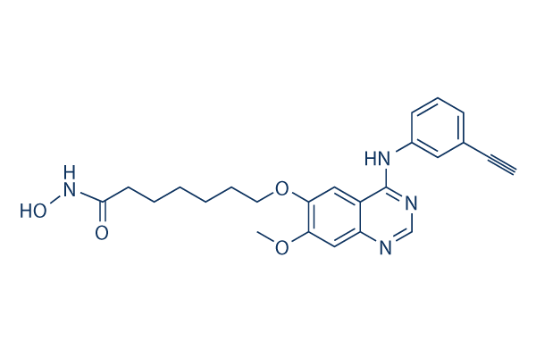
Estructura química
Peso molecular: 434.49
Control de calidad
Cultivo celular, tratamiento y concentración de trabajo
| Líneas celulares | Tipo de ensayo | Concentración | Tiempo de incubación | Formulación | Descripción de la actividad | PMID |
|---|---|---|---|---|---|---|
| human SK-BR-3 cells | Proliferation assay | Antiproliferative activity against human SK-BR-3 cells after hrs by ATP content assay, IC50=0.04 μM | ||||
| MDA-MB-231 cells | Proliferation assay | Antiproliferative activity against human MDA-MB-231 cells after hrs by ATP content assay, IC50=0.1 μM | ||||
| human HepG2 cells | Proliferation assay | Antiproliferative activity against human HepG2 cells after hrs by ATP content assay, IC50=0.13 μM | ||||
| human SKHEP1 cells | Proliferation assay | Antiproliferative activity against human SKHEP1 cells after hrs by ATP content assay, IC50=0.22 μM | ||||
| human Hep3B2 cells | Proliferation assay | Antiproliferative activity against human Hep3B2 cells after hrs by ATP content assay, IC50=0.23 μM | ||||
| human BxPC3 cells | Proliferation assay | Antiproliferative activity against human BxPC3 cells after hrs by ATP content assay, IC50=0.27 μM | ||||
| human NCI-H358 cells | Proliferation assay | Antiproliferative activity against human NCI-H358 cells after hrs by ATP content assay, IC50=0.4 μM | ||||
| human MCF7 cells | Proliferation assay | Antiproliferative activity against human MCF7 cells after hrs by ATP content assay, IC50=0.55 μM | ||||
| human HCC827 cells | Proliferation assay | Antiproliferative activity against human HCC827 cells after hrs by ATP content assay, IC50=0.6 μM | ||||
| human H460 cells | Proliferation assay | Antiproliferative activity against human H460 cells after hrs by ATP content assay, IC50=0.7 μM | ||||
| human Capan1 cells | Proliferation assay | Antiproliferative activity against human Capan1 cells after hrs by ATP content assay, IC50=0.8 μM | ||||
| Haga clic para ver más datos experimentales de líneas celulares | ||||||
Información química, almacenamiento y estabilidad
| Peso molecular | 434.49 | Fórmula | C24H26N4O4 |
Almacenamiento (Desde la fecha de recepción) | |
|---|---|---|---|---|---|
| Nº CAS | 1012054-59-9 | Descargar SDF | Almacenamiento de soluciones madre |
|
|
| Sinónimos | N/A | Smiles | COC1=C(C=C2C(=C1)N=CN=C2NC3=CC=CC(=C3)C#C)OCCCCCCC(=O)NO | ||
Solubilidad
|
In vitro |
DMSO
: 43 mg/mL
(98.96 mM)
Water : Insoluble Ethanol : Insoluble |
Calculadora de Molaridad
|
In vivo |
|||||
Calculadora de formulación in vivo (Solución clara)
Paso 1: Introduzca la información a continuación (Recomendado: Un animal adicional para tener en cuenta la pérdida durante el experimento)
Paso 2: Introduzca la formulación in vivo (Esto es solo la calculadora, no la formulación. Por favor, contáctenos primero si no hay una formulación in vivo en la sección de Solubilidad.)
Resultados del cálculo:
Concentración de trabajo: mg/ml;
Método para preparar el líquido maestro de DMSO: mg fármaco predissuelto en μL DMSO ( Concentración del líquido maestro mg/mL, Por favor, contáctenos primero si la concentración excede la solubilidad del DMSO del lote del fármaco. )
Método para preparar la formulación in vivo: Tomar μL DMSO líquido maestro, luego añadirμL PEG300, mezclar y clarificar, luego añadirμL Tween 80, mezclar y clarificar, luego añadir μL ddH2O, mezclar y clarificar.
Método para preparar la formulación in vivo: Tomar μL DMSO líquido maestro, luego añadir μL Aceite de maíz, mezclar y clarificar.
Nota: 1. Por favor, asegúrese de que el líquido esté claro antes de añadir el siguiente disolvente.
2. Asegúrese de añadir el (los) disolvente(s) en orden. Debe asegurarse de que la solución obtenida, en la adición anterior, sea una solución clara antes de proceder a añadir el siguiente disolvente. Se pueden utilizar métodos físicos como el vórtice, el ultrasonido o el baño de agua caliente para ayudar a la disolución.
Mecanismo de acción
| Targets/IC50/Ki |
EGFR
(Cell-free assay) 2.4 nM
HDAC
(Cell-free assay) 4.4 nM
HDAC1
(Cell-free assay) 4.5 nM
HDAC6
(Cell-free assay) 5.1 nM
HDAC3
(Cell-free assay) 9.1 nM
HDAC5
(Cell-free assay) 11.4 nM
HDAC2
(Cell-free assay) 12.6 nM
HDAC4
(Cell-free assay) 13.2 nM
HER2
(Cell-free assay) 15.7 nM
HDAC10
(Cell-free assay) 26.1 nM
HDAC9
(Cell-free assay) 67.2 nM
HDAC8
(Cell-free assay) 79.8 nM
HDAC7
(Cell-free assay) 373 nM
|
|---|---|
| In vitro |
Específico para las HDAC de clase I y clase II, CUDC-101 no inhibe las HDAC de tipo Sir de clase III. Este compuesto muestra una actividad débil contra otras proteínas quinasas, incluyendo KDR/VEGFR2, Lyn, Lck, Abl-1, FGFR-2, Flt-3 y Ret con IC50 de 0.85 μM, 0.84 μM, 5.91 μM, 2.89 μM, 3.43 μM, 1.5 μM y 3.2 μM, respectivamente. Muestra una amplia actividad antiproliferativa en muchos tipos de células cancerosas humanas con IC50 de 0.04-0.80 μM, exhibiendo una mayor potencia y combinaciones de vorinostat en la mayoría de los casos. Este químico inhibe potentemente las líneas celulares cancerosas. Inhibe la mutante T790M de EGFR resistente aunque sus efectos son incompletos con un Amax de ~60% de la actividad enzimática máxima después de la inhibición. El tratamiento con este compuesto aumenta la acetilación de las histonas H3 y H4, así como la acetilación de sustratos no histonas de HDAC como p53 y α-tubulina, de manera dosis-dependiente en varias líneas celulares cancerosas. También suprime la expresión de HER3, la amplificación de Met y la reactivación de AKT en células tumorales. |
| Ensayo de quinasa |
Ensayos de inhibición de HDAC, EGFR y HER2
|
|
Las actividades de las HDAC de Clase I y II se evalúan utilizando el sistema Biomol Color de Lys. En resumen, los extractos nucleares de células HeLa se utilizan como fuente de HDAC. Se añaden diferentes concentraciones de CUDC-101 a los extractos nucleares de células HeLa en presencia de un sustrato artificial colorimétrico. Se añade revelador al final del ensayo y la actividad enzimática se mide en el lector de microplacas Wallac Victor II 1420 a 405 nM. La actividad quinasa de EGFR y HER2 se mide utilizando los kits de ensayo de quinasa HTScan EGF receptor y HER2. En resumen, la proteína de fusión GST-EGFR se incuba con un péptido sustrato biotinilado sintético y varias concentraciones de este compuesto en presencia de 400 mM de ATP. El sustrato fosforilado se captura con placas de 96 pocillos recubiertas de estreptavidina. El nivel de fosforilación se monitoriza mediante anticuerpos secundarios anti-fosfotirosina y marcados con europio. La solución de mejora se añade al final del ensayo y la actividad enzimática se mide en el lector de microplacas Wallac Victor II 1420 a 615 nM.
|
|
| In vivo |
La administración de CUDC-101 a 120 mg/kg/día induce la regresión tumoral en el modelo de cáncer de hígado Hep-G2, que es más eficaz a su dosis máxima tolerada (25 mg/kg/día) y vorinostat a una dosis de concentración equimolar (72 mg/kg/día). Este compuesto inhibe el crecimiento de xenoinjertos de CPNM H358 sensibles de manera dosis-dependiente. También muestra una potente inhibición del crecimiento tumoral en el modelo de xenoinjerto de CPNM A549 resistente. Este químico produce una regresión tumoral significativa en el modelo de cáncer de mama MDA-MB-468 resistente, HER2-negativo, con sobreexpresión de EGFR y el modelo de carcinoma de células escamosas de cabeza y cuello (HNSCC) CAL-27 con sobreexpresión de EGFR. Además, inhibe el crecimiento tumoral en los modelos de cáncer colorrectal HCT116 mutante K-ras y de cáncer de páncreas HPAC con expresión de EGFR/HER2 (neu). |
Referencias |
|
Información del ensayo clínico
(datos de https://clinicaltrials.gov, actualizado el 2024-05-22)
| Número NCT | Reclutamiento | Condiciones | Patrocinador/Colaboradores | Fecha de inicio | Fases |
|---|---|---|---|---|---|
| NCT01702285 | Terminated | Cancer |
Curis Inc. |
September 2012 | Phase 1 |
| NCT01384799 | Completed | Head and Neck Cancer |
Curis Inc. |
November 2011 | Phase 1 |
| NCT00728793 | Completed | Tumors |
Curis Inc. |
August 2008 | Phase 1 |
Soporte técnico
Tel: +1-832-582-8158 Ext:3
Si tiene alguna otra consulta, por favor deje un mensaje.

Los productos son solo para uso de investigación. No para uso humano. No vendemos a pacientes.
©Copyright 2013 Selleck Chemicals. Todos los derechos reservados.