
solo para uso en investigación
Menadione (Vitamin K3) phosphatase inhibidor
Cat. No.S1949
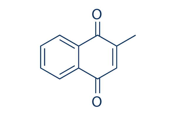
Estructura química
Peso molecular: 172.18
Control de calidad
Cultivo celular, tratamiento y concentración de trabajo
| Líneas celulares | Tipo de ensayo | Concentración | Tiempo de incubación | Formulación | Descripción de la actividad | PMID |
|---|---|---|---|---|---|---|
| HT22 cells | Function assay | Neuroprotective activity in mouse HT22 cells assessed as prevention from glutamate-induced oxidative cell death by MTS assay, PD50=0.797 μM | ||||
| HeLa cell | Cytotoxicity assay | Cytotoxicity against HeLa cell lines, EC50=3.7 μM | ||||
| PaCa2 cell | Proliferation assay | Antiproliferative activity against human pancreatic carcinoma Mia PaCa2 cell line by colorimetric assay, IC50=6.2 μM | ||||
| HT-29 cells | Cytotoxicity assay | 72 h | Cytotoxicity against human HT-29 cells assessed as growth inhibition after 72 hrs by MTS assay, IC50=9.69 μM | |||
| DU145 cells | Cytotoxicity assay | 72 h | Cytotoxicity against human DU145 cells assessed as growth inhibition after 72 hrs by MTS assay, IC50=9.86 μM | |||
| MDA-MB-231 cells | Cytotoxicity assay | 72 h | Cytotoxicity against human MDA-MB-231 cells assessed as growth inhibition after 72 hrs by MTS assay, IC50=12.68 μM | |||
| human A2058 cells | Cytotoxicity assay | Cytotoxicity against human A2058 cells assessed as inhibition of formazan formation at day 5 by measuring absorbance at 430 nm, IC50=13.4 μM | ||||
| SAS cells | Cytotoxicity assay | 48 h | Cytotoxicity against human SAS cells assessed as reduction in cell viability after 48 hrs by MTT assay, IC50=14 μM | |||
| HaCaT cells | Function assay | 48 h | Antihyperproliferative activity against human HaCaT cells assessed as inhibition of cell growth after 48 hrs by phase contrast microscopy, IC50=15.8 μM | |||
| HeLa cells | Cytotoxicity assay | 48 h | Cytotoxicity against human HeLa cells assessed as reduction in cell viability after 48 hrs by MTT assay, IC50=18 μM | |||
| CRL2796 cells | Cytotoxicity assay | Cytotoxicity against human CRL2796 cells assessed as inhibition of formazan formation at day 4 by measuring absorbance at 430 nm, IC50=21 μM | ||||
| HL60 cells | Function assay | 50 μM | Induction of necrosis in intact human HL60 cells assessed as cytosolic LDH release at 50 uM | |||
| Haga clic para ver más datos experimentales de líneas celulares | ||||||
Información química, almacenamiento y estabilidad
| Peso molecular | 172.18 | Fórmula | C11H8O2 |
Almacenamiento (Desde la fecha de recepción) | |
|---|---|---|---|---|---|
| Nº CAS | 58-27-5 | Descargar SDF | Almacenamiento de soluciones madre |
|
|
| Sinónimos | NSC 4170 | Smiles | CC1=CC(=O)C2=CC=CC=C2C1=O | ||
Solubilidad
|
In vitro |
DMSO
: 34 mg/mL
(197.46 mM)
Ethanol : 34 mg/mL Water : Insoluble |
Calculadora de Molaridad
|
In vivo |
|||||
Calculadora de formulación in vivo (Solución clara)
Paso 1: Introduzca la información a continuación (Recomendado: Un animal adicional para tener en cuenta la pérdida durante el experimento)
Paso 2: Introduzca la formulación in vivo (Esto es solo la calculadora, no la formulación. Por favor, contáctenos primero si no hay una formulación in vivo en la sección de Solubilidad.)
Resultados del cálculo:
Concentración de trabajo: mg/ml;
Método para preparar el líquido maestro de DMSO: mg fármaco predissuelto en μL DMSO ( Concentración del líquido maestro mg/mL, Por favor, contáctenos primero si la concentración excede la solubilidad del DMSO del lote del fármaco. )
Método para preparar la formulación in vivo: Tomar μL DMSO líquido maestro, luego añadirμL PEG300, mezclar y clarificar, luego añadirμL Tween 80, mezclar y clarificar, luego añadir μL ddH2O, mezclar y clarificar.
Método para preparar la formulación in vivo: Tomar μL DMSO líquido maestro, luego añadir μL Aceite de maíz, mezclar y clarificar.
Nota: 1. Por favor, asegúrese de que el líquido esté claro antes de añadir el siguiente disolvente.
2. Asegúrese de añadir el (los) disolvente(s) en orden. Debe asegurarse de que la solución obtenida, en la adición anterior, sea una solución clara antes de proceder a añadir el siguiente disolvente. Se pueden utilizar métodos físicos como el vórtice, el ultrasonido o el baño de agua caliente para ayudar a la disolución.
Mecanismo de acción
| Targets/IC50/Ki |
Cdc25 phosphatase
DNA polymerase γ
|
|---|---|
| In vitro |
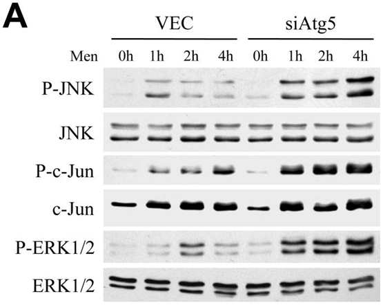
La Menadione (Vitamin K3) previene la captación mitocondrial de Ca(2+) en las células acinares pancreáticas normales, lo que permite una rápida propagación del Ca(2+) por toda la célula. Su despolarización mitocondrial inducida se debe a la inducción del poro de transición de permeabilidad. A concentraciones no tóxicas, este compuesto produce una breve activación de la quinasa regulada por señal extracelular (ERK) y la quinasa N-terminal c-Jun (JNK) en la línea celular de hepatocitos de rata RALA255-10G. Es equipotente contra líneas celulares de leucemia multirresistentes y parentales con valores de IC50 de 13,5 mM y 18 mM, respectivamente. El compuesto inhibe la incorporación de timidina, uridina y aminoácidos radiactivos en el ADN, ARN y proteínas, respectivamente, en tres líneas celulares de cáncer humano. A 1-20 mM, inhibe la proliferación celular de las células AR4-2J de forma dosis- y tiempo-dependiente. Una concentración de 100 mM causa una muerte celular rápida. Este nivel (100 mM) induce un frotis de ADN en la electroforesis indicativo de necrosis, mientras que concentraciones más bajas (10-20 mM) inducen una escalera de ADN indicativa de apoptosis. Las concentraciones de 1-20 mM inducen el P53 de tipo salvaje, mientras que 100 mM tiene un efecto menor sobre el P53 de tipo salvaje. El tratamiento con este compuesto afecta claramente la función mitocondrial de las células T Jurkat al inducir un colapso del potencial transmembrana interno (DeltaPsi(m)) y una disminución de la masa de la membrana interna, que podría revertirse completamente con N-acetilcisteína. |
Referencias |
|
Aplicaciones
| Métodos | Biomarcadores | Imágenes | PMID |
|---|---|---|---|
| Western blot | p-JNK / JNK / p-c-Jun / c-Jun / p-ERK / ERK Cyclin B1 / CDK1 / CDC25C c-Fos / HO-1 |
|
20578144 |
Información del ensayo clínico
(datos de https://clinicaltrials.gov, actualizado el 2024-05-22)
| Número NCT | Reclutamiento | Condiciones | Patrocinador/Colaboradores | Fecha de inicio | Fases |
|---|---|---|---|---|---|
| NCT01094444 | Completed | Folliculitis |
Per Pfeiffer|Odense University Hospital |
May 2010 | Phase 2 |
| NCT00656786 | Completed | EGFR Inhibitor-associated Rash |
Spectrum Pharmaceuticals Inc |
April 3 2008 | Phase 1 |
Soporte técnico
Tel: +1-832-582-8158 Ext:3
Si tiene alguna otra consulta, por favor deje un mensaje.

Los productos son solo para uso de investigación. No para uso humano. No vendemos a pacientes.
©Copyright 2013 Selleck Chemicals. Todos los derechos reservados.