solo para uso en investigación
Berberine chloride Anti-infection químico
Cat. No.S2271
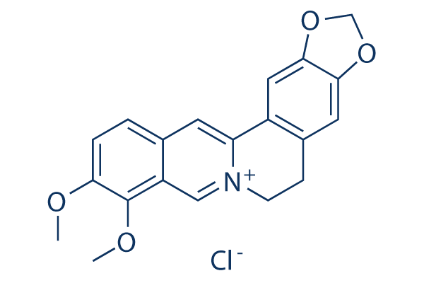
Estructura química
Peso molecular: 371.81
Control de calidad
Cultivo celular, tratamiento y concentración de trabajo
| Líneas celulares | Tipo de ensayo | Concentración | Tiempo de incubación | Formulación | Descripción de la actividad | PMID |
|---|---|---|---|---|---|---|
| MRC5 cells | Function assay | Antiviral activity against HCMV in MRC5 cells by plaque reduction assay, IC50=0.68 μM | ||||
| MRC5 cells | Proliferation assay | 10 μM | 54 hrs | Inhibition of HCMV proliferation in MRC5 cells after 54 hrs post-infection at 10 uM by plaque assay | ||
| MRC5 cells | Proliferation assay | 10 μM | 24 h | Inhibition of HCMV proliferation in MRC5 cells after 24 hrs post-infection at 10 uM by plaque assay | ||
| Bel7402 cells | Function assay | 12 h | Induction of LDLR protein in human Bel7402 cells after 12 hrs by RT-PCR assay relative to control | |||
| HepG2 cells | Function assay | 10 ug/mL | 12 h | Induction of LDLR protein expression in human HepG2 cells at 10 ug/mL after 12 hrs by flow cytometry | ||
| KB cells | Cytotoxicity assay | 72 h | Cytotoxicity against human KB cells after 72 hrs, IC50=7.32 μM | |||
| HL60 cells | Apoptosis assay | 48 hrs | Induction of apoptosis in human HL60 cells after 48 hrs using annexin V-propidium iodide staining by FACS analysis | |||
| A549 cells | Cytotoxicity assay | Cytotoxicity against human A549 cells by SRB assay, IC50=6.27 μM | ||||
| SKOV3 cells | Cytotoxicity assay | Cytotoxicity against human SKOV3 cells by SRB assay, IC50=16.44 μM | ||||
| SK-MEL-2 cells | Cytotoxicity assay | Cytotoxicity against human SK-MEL-2 cells by SRB assay, IC50=13.76 μM | ||||
| HCT15 cells | Cytotoxicity assay | Cytotoxicity against human HCT15 cells by SRB assay, IC50=16.59 μM | ||||
| CEM cells | Cytotoxicity assay | 48 hrs | Cytotoxicity against human CEM cells expressing green fluorescent protein after 48 hrs by MTT assay, CC50=2.09 μM | |||
| human CEM cells | Function assay | 7 days | Antiviral activity against 0.05 MOI Human immunodeficiency virus 1 NL4.3 infected in human CEM cells expressing green fluorescent protein assessed as p24 antigen production measured 7 days post infection by ELISA, EC50=0.13 μM | |||
| SKN cells | Growth inhibition assay | 72 h | Growth inhibition against human SKN cells after 72 hrs by MTT assay, GI50=15.88 μM | |||
| RKN cells | Growth inhibition assay | 48 hrs | Growth inhibition against human RKN cells after 48 hrs by MTT assay, GI50=49.6 μM | |||
| G402 cells | Growth inhibition assay | 48 hrs | Growth inhibition against human G402 cells after 48 hrs by MTT assay, GI50=11.87 μM | |||
| A10 cells | Function assay | 30 μM | 24 hrs | Downregulation of Scd2 mRNA expression in rat A10 cells at 30 uM after 24 hrs by quantitative RT-PCR analysis | ||
| A10 cells | Function assay | 30 μM | 24 hrs | Down regulation of Prim2 mRNA expression in rat A10 cells at 30 uM after 24 hrs by quantitative RT-PCR analysis | ||
| A10 cells | Function assay | 30 μM | 24 hrs | Downregulation of Impk mRNA expression in rat A10 cells at 30 uM after 24 hrs by quantitative RT-PCR analysis | ||
| HepG2-A16-CD81 cells | Function assay | 10 μM | NOVARTIS: Antimalarial liver stage activity measured as a greater than 50% reduction in Plasmodium yoelii schizont area in HepG2-A16-CD81 cells at 10uM compound concentration, determined by immuno-fluorescence. | |||
| HepG2-A16-CD81 cells | Function assay | 10 μM | NOVARTIS: Antimalarial liver stage activity measured as reduction in Plasmodium yoelii schizont area in HepG2-A16-CD81 cells by immuno-fluorescence, and median schizont size at 10uM compound concentration, IC50=0.548 μM | |||
| HepG2 cells | Function assay | 10 μM | 4 h | Increase in AMPKalpha phosphorylation in human HepG2 cells at 10 uM after 4 hrs by Western blot analysis relative to untreated control | ||
| HepG2 cells | Function assay | 10 μM | 4 h | Increase in total AMPKalpha level in human HepG2 cells at 10 uM after 4 hrs by Western blot analysis relative to untreated control | ||
| HepG2 cells | Function assay | 20 μM | 24 hrs | Induction of apoptosis in human HepG2 cells assessed as morphological changes at 20 uM after 24 hrs using Hoechst 33258 staining by fluorescence microscopic analysis | ||
| HT-29 cells | Cytotoxicity assay | 48 hrs | Cytotoxicity against human HT-29 cells after 48 hrs by MTT assay, IC50=8.45 μM | |||
| HepG2 cells | Cytotoxicity assay | 24 hrs | Cytotoxicity against human HepG2 cells after 24 hrs by MTT assay, IC50=11.22 μM | |||
| HepG2 cells | Cytotoxicity assay | 48 hrs | Cytotoxicity against human HepG2 cells after 48 hrs by MTT assay, IC50=8.32 μM | |||
| Haga clic para ver más datos experimentales de líneas celulares | ||||||
Información química, almacenamiento y estabilidad
| Peso molecular | 371.81 | Fórmula | C20H18NO4.Cl |
Almacenamiento (Desde la fecha de recepción) | |
|---|---|---|---|---|---|
| Nº CAS | 633-65-8 | Descargar SDF | Almacenamiento de soluciones madre |
|
|
Solubilidad
|
In vitro |
DMSO
: 25 mg/mL
(67.23 mM)
Water : Insoluble Ethanol : Insoluble |
Calculadora de Molaridad
|
In vivo |
|||||
Calculadora de formulación in vivo (Solución clara)
Paso 1: Introduzca la información a continuación (Recomendado: Un animal adicional para tener en cuenta la pérdida durante el experimento)
Paso 2: Introduzca la formulación in vivo (Esto es solo la calculadora, no la formulación. Por favor, contáctenos primero si no hay una formulación in vivo en la sección de Solubilidad.)
Resultados del cálculo:
Concentración de trabajo: mg/ml;
Método para preparar el líquido maestro de DMSO: mg fármaco predissuelto en μL DMSO ( Concentración del líquido maestro mg/mL, Por favor, contáctenos primero si la concentración excede la solubilidad del DMSO del lote del fármaco. )
Método para preparar la formulación in vivo: Tomar μL DMSO líquido maestro, luego añadirμL PEG300, mezclar y clarificar, luego añadirμL Tween 80, mezclar y clarificar, luego añadir μL ddH2O, mezclar y clarificar.
Método para preparar la formulación in vivo: Tomar μL DMSO líquido maestro, luego añadir μL Aceite de maíz, mezclar y clarificar.
Nota: 1. Por favor, asegúrese de que el líquido esté claro antes de añadir el siguiente disolvente.
2. Asegúrese de añadir el (los) disolvente(s) en orden. Debe asegurarse de que la solución obtenida, en la adición anterior, sea una solución clara antes de proceder a añadir el siguiente disolvente. Se pueden utilizar métodos físicos como el vórtice, el ultrasonido o el baño de agua caliente para ayudar a la disolución.
Mecanismo de acción
| Targets/IC50/Ki |
Caspase-3
Caspase-8
PARP
cytochrome c
cIAP1
Bcl-2
Bcl-xL
JNK
p38 MAPK
ROS
Topo I
Topo II
|
|---|---|
| In vitro |
En comparación con el regorafenib solo, el tratamiento combinado de Berberine (BBR) y regorafenib inhibe significativamente la proliferación de células de carcinoma hepatocelular (HCC) e induce la apoptosis celular. |
| In vivo |
El grupo de tratamiento combinado con Berberine (BBR) y regorafenib tiene un efecto inhibidor dramático sobre el crecimiento de tumores de xenoinjerto de carcinoma hepatocelular (HCC) en ratones desnudos. Se observa un aumento de la apoptosis de los tumores de xenoinjerto en el grupo de tratamiento combinado. |
Referencias |
|
Información del ensayo clínico
(datos de https://clinicaltrials.gov, actualizado el 2024-05-22)
| Número NCT | Reclutamiento | Condiciones | Patrocinador/Colaboradores | Fecha de inicio | Fases |
|---|---|---|---|---|---|
| NCT06273241 | Not yet recruiting | Pharmacokinetic Study in Healthy Volunteers |
University Medicine Greifswald |
March 4 2024 | Not Applicable |
| NCT05845931 | Recruiting | Pharmacokinetic Study in Healthy Volunteers |
University Medicine Greifswald |
May 5 2023 | Not Applicable |
| NCT05480670 | Completed | Polycystic Ovary Syndrome |
Ayub Teaching Hospital |
November 1 2022 | Not Applicable |
| NCT05463003 | Completed | Pharmacokinetic Study in Healthy Volunteers |
University Medicine Greifswald |
July 19 2022 | Not Applicable |
Soporte técnico
Tel: +1-832-582-8158 Ext:3
Si tiene alguna otra consulta, por favor deje un mensaje.
Los productos son solo para uso de investigación. No para uso humano. No vendemos a pacientes.
©Copyright 2013 Selleck Chemicals. Todos los derechos reservados.