Solo para uso en investigación
Melanoma Associated Antigen 100+ / 7 kDa Antibody [G24P23]
Nº de cat.: F3175
Aplicación:
Reactividad:
-
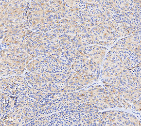 Immunohistochemical analysis of formalin fixed paraffin embedded human melanoma tissue with F3175 at 1:20 dilution.
Información de uso
| Dilución |
|---|
|
| Aplicación |
|---|
| IHC, FCM |
| Reactividad |
|---|
| Human |
| Fuente |
|---|
| Mouse Monoclonal Antibody |
| Tampón de almacenamiento |
|---|
| PBS, pH 7.2+50% Glycerol+0.05% BSA+0.01% NaN3 |
| Almacenamiento (desde la fecha de recepción) |
|---|
| -20°C (avoid freeze-thaw cycles), 2 years |
| Control positivo | Human melanoma; Malme-3 |
|---|---|
| Control negativo |
Métodos experimentales
| IHC |
|---|
Experimental Protocol:
Deparaffinization/Rehydration
1. Deparaffinize/hydrate sections:
2. Incubate sections in three washes of xylene for 5 min each.
3. Incubate sections in two washes of 100% ethanol for 10 min each.
4. Incubate sections in two washes of 95% ethanol for 10 min each.
5. Wash sections two times in dH2O for 5 min each.
6.Antigen retrieval: For Citrate: Heat slides in a microwave submersed in 1X citrate unmasking solution until boiling is initiated; continue with 10 min at a sub-boiling temperature (95°-98°C). Cool slides on bench top for 30 min.
Staining
1. Wash sections in dH2O three times for 5 min each.
2. Incubate sections in 3% hydrogen peroxide for 10 min.
3. Wash sections in dH2O two times for 5 min each.
4. Wash sections in wash buffer for 5 min.
5. Block each section with 100–400 µl of blocking solution for 1 hr at room temperature.
6. Remove blocking solution and add 100–400 µl primary antibody diluent in to each section. Incubate overnight at 4°C.
7. Remove antibody solution and wash sections with wash buffer three times for 5 min each.
8. Cover section with 1–3 drops HRPas needed. Incubate in a humidified chamber for 30 min at room temperature.
9. Wash sections three times with wash buffer for 5 min each.
10. Add DAB Chromogen Concentrate to DAB Diluent and mix well before use.
11. Apply 100–400 µl DAB to each section and monitor closely. 1–10 min generally provides an acceptable staining intensity.
12. Immerse slides in dH2O.
13. If desired, counterstain sections with hematoxylin.
14. Wash sections in dH2O two times for 5 min each.
15. Dehydrate sections: Incubate sections in 95% ethanol two times for 10 sec each; Repeat in 100% ethanol, incubating sections two times for 10 sec each; Repeat in xylene, incubating sections two times for 10 sec each.
16. Mount sections with coverslips and mounting medium.
|
Descripción biológica
| Especificidad |
|---|
Melanoma Associated Antigen 100+ / 7 kDa Antibody [G24P23] detects endogenous levels of total Melanoma Associated Antigen 100+ / 7 kDa protein. |
| Localización subcelular |
|---|
| Endoplasmic reticulum, Endosome, Golgi apparatus, Membrane, Secreted |
| Uniprot ID |
|---|
| P40967 |
| Clon |
|---|
| G24P23 |
| Sinónimo |
|---|
| MAA |
| Antecedentes |
|---|
| Melanoma Associated Antigen 100+ / 7 kDa (MAGE) proteins are tumor-associated antigens, typically restricted to germ cells of the testis in normal adult tissues but aberrantly expressed in various cancers, including melanoma, glioma, lung, bladder, and multiple myeloma. The “100+/7 kDa” designation refers to variants or isoforms with molecular weights around 100 kDa and 7 kDa. Structurally, MAGE proteins share a conserved ~200 amino acid MAGE homology domain (MHD) composed of two winged helix domains forming a deep cleft for peptide or protein binding, flanked by variable N- and C-terminal regions. Functionally, MAGE proteins can act as oncogenic drivers by interacting with RING-type E3 ubiquitin ligases, modulating protein degradation pathways that regulate apoptosis, proliferation, and metabolism; for example, MAGE-A3 promotes tumor survival by stimulating TRIM28-mediated p53 degradation, whereas MAGE-A4 may induce apoptosis via different protein partners. Their tumor-specific expression, structural plasticity, and oncogenic roles make MAGEs both biomarkers and potential therapeutic targets in cancer immunotherapy. |
| Referencias |
|---|
|
Soporte técnico
Tel: +1-832-582-8158 Ext:3
Si tiene alguna otra consulta, por favor deje un mensaje.
Los productos son solo para uso de investigación. No para uso humano. No vendemos a pacientes.
©Copyright 2013 Selleck Chemicals. Todos los derechos reservados.