Solo para uso en investigación
Phospho-PRC1 (Thr481) Antibody [H23N3]
Nº de cat.: F3675
Aplicación:
Reactividad:
-
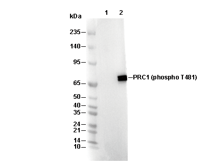 Lane 1: Hela, Lane 2: Hela (aphidicolin + nocodazole treated)
Información de uso
| Dilución |
|---|
|
| Aplicación |
|---|
| WB |
| Reactividad |
|---|
| Human |
| Fuente |
|---|
| Rabbit Monoclonal Antibody |
| Tampón de almacenamiento |
|---|
| PBS, pH 7.2+50% Glycerol+0.05% BSA+0.01% NaN3 |
| Almacenamiento (desde la fecha de recepción) |
|---|
| -20°C (avoid freeze-thaw cycles), 2 years |
| PM predicho |
|---|
| 72 kDa |
| Control positivo | HeLa cell (treated with aphidicolin and nocodazole) |
|---|---|
| Control negativo |
Métodos experimentales
| WB |
|---|
Experimental Protocol:
Sample preparation
1. Tissue: Lyse the tissue sample by adding an appropriate volume of ice-cold RIPA/NP-40 Lysis Buffer (containing Protease Inhibitor Cocktail, Phosphatase Inhibitor Cocktail),and homogenize the tissue at a low temperature. 2. Adherent cell: Aspirate the culture medium and wash the cells with ice-cold PBS twice. Lyse the cells by adding an appropriate volume of RIPA/NP-40 Lysis Buffer (containing Protease Inhibitor Cocktail, Phosphatase Inhibitor Cocktail) and put the sample on ice for 5 min. 3. Suspension cell: Transfer the culture medium to a pre-cooled centrifuge tube. Centrifuge and aspirate the supernatant. Wash the cells with ice-cold PBS twice. Lyse the cells by adding an appropriate volume of RIPA/NP-40 Lysis Buffer (containing Protease Inhibitor Cocktail, Phosphatase Inhibitor Cocktail) and put the sample on ice for 5 min. 4. Place the lysate into a pre-cooled microcentrifuge tube. Centrifuge at 4°C for 15 min. Collect the supernatant;
5. Remove a small volume of lysate to determine the protein concentration;
6. Combine the lysate with protein loading buffer. Boil 20 µL sample under 95-100°C for 5 min. Centrifuge for 5 min after cool down on ice.
Electrophoretic separation
1. According to the concentration of extracted protein, load appropriate amount of protein sample and marker onto SDS-PAGE gels for electrophoresis. Recommended separating gel (lower gel) concentration: 10%. Reference Table for Selecting SDS-PAGE Separation Gel Concentrations 2. Power up 80V for 30 minutes. Then the power supply is adjusted (110 V~150 V), the Marker is observed, and the electrophoresis can be stopped when the indicator band of the predyed protein Marker where the protein is located is properly separated. (Note that the current should not be too large when electrophoresis, too large current (more than 150 mA) will cause the temperature to rise, affecting the result of running glue. If high currents cannot be avoided, an ice bath can be used to cool the bath.)
Transfer membrane
1. Take out the converter, soak the clip and consumables in the pre-cooled converter;
2. Activate PVDF membrane with methanol for 1 min and rinse with transfer buffer;
3. Install it in the order of "black edge of clip - sponge - filter paper - filter paper - glue -PVDF membrane - filter paper - filter paper - sponge - white edge of clip"; 4. The protein was electrotransferred to PVDF membrane. ( 0.45 µm PVDF membrane is recommended ) Reference Table for Selecting PVDF Membrane Pore Size Specifications Recommended conditions for wet transfer: 200 mA, 120 min. ( Note that the transfer conditions can be adjusted according to the protein size. For high-molecular-weight proteins, a higher current and longer transfer time are recommended. However, ensure that the transfer tank remains at a low temperature to prevent gel melting.)
Block
1. After electrotransfer, wash the film with TBST at room temperature for 5 minutes;
2. Incubate the film in the blocking solution ( recommending 5% BSA solution)
for 1 hour at room temperature;
3. Wash the film with TBST for 3 times, 5 minutes each time.
Antibody incubation
1. Use 5% skim milk powder to prepare the primary antibody working liquid (recommended dilution ratio for primary antibody 1:500), gently shake and incubate with the film at 4°C overnight; 2. Wash the film with TBST 3 times, 5 minutes each time;
3. Add the secondary antibody to the blocking solution and incubate with the film gently at room temperature for 1 hour;
4. After incubation, wash the film with TBST 3 times for 5 minutes each time.
Antibody staining
1. Add the prepared ECL luminescent substrate (or select other color developing substrate according to the second antibody) and mix evenly;
2. Incubate with the film for 1 minute, remove excess substrate (keep the film moist), wrap with plastic film, and expose in the imaging system. |
Descripción biológica
| Especificidad |
|---|
Phospho-PRC1 (Thr481) Antibody [H23N3] recognizes endogenous levels of total PRC1 protein only when phosphorylated at Thr 481. |
| Localización subcelular |
|---|
| Chromosome, Cytoplasm, Cytoskeleton, Microtubule, Nucleus |
| Uniprot ID |
|---|
| O43663 |
| Clon |
|---|
| H23N3 |
| Sinónimo |
|---|
| Protein regulator of cytokinesis 1, PRC1 |
| Antecedentes |
|---|
| Phospho-PRC1 (Thr481) refers to the phosphorylated form of the microtubule-binding protein PRC1 at threonine 481, a key regulatory site within its C-terminal region. PRC1 is crucial for bundling antiparallel microtubules at the spindle midzone during cell division, and its activity is tightly regulated: during early mitosis, Cdk1/cyclin B phosphorylates PRC1 at Thr481, inhibiting its bundling function to prevent premature spindle stabilization; as cells progress to anaphase and Cdk1 activity declines, dephosphorylation at Thr481 reactivates PRC1, enabling proper spindle midzone formation and successful cytokinesis. PRC1 is expressed at high levels during S and G2/M phases, localizes dynamically from the nucleus to the spindle and midbody, and forms homodimers that crosslink microtubules, collaborating with proteins like Kinesin-4 to organize the central spindle. Dysregulation of PRC1 phosphorylation or expression can disrupt spindle assembly, block cytokinesis, and contribute to chromosomal instability and tumor progression |
| Referencias |
|---|
|
Soporte técnico
Tel: +1-832-582-8158 Ext:3
Si tiene alguna otra consulta, por favor deje un mensaje.
Los productos son solo para uso de investigación. No para uso humano. No vendemos a pacientes.
©Copyright 2013 Selleck Chemicals. Todos los derechos reservados.