Solo para uso en investigación
Phospho-GSK-3α/β (Ser21/9) Antibody [N3H2]
Nº de cat.: F0496
Aplicación:
Reactividad:
-
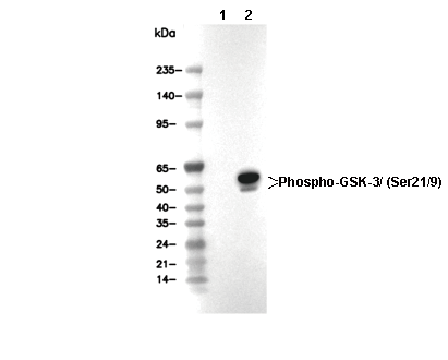 Lane 1: COS-7 (λ phosphatase treated), Lane 2: COS-7 (PDGF, 100ng/mL, 5 min)
Información de uso
| Dilución |
|---|
|
| Aplicación |
|---|
| WB, IP |
| Reactividad |
|---|
| Human, Mouse, Rat, Monkey |
| Fuente |
|---|
| Rabbit Monoclonal Antibody |
| Tampón de almacenamiento |
|---|
| PBS, pH 7.2+50% Glycerol+0.05% BSA+0.01% NaN3 |
| Almacenamiento (desde la fecha de recepción) |
|---|
| -20°C (avoid freeze-thaw cycles), 2 years |
| PM predicho |
|---|
| 51 kDa, 46 kDa |
| Control positivo | COS-7 cell (PDGF-treated); C6 cell; 293 cell (Insulin, 100nM, 20 min); NIH/3T3 cell (PDGF, 100ng/mL, 5 min); HeLa cell |
|---|---|
| Control negativo | COS-7 cell; C6 cell (PDGF, 100ng/mL, 5 min); 293 cell; NIH/3T3 cell |
Métodos experimentales
| WB |
|---|
Experimental Protocol:
Sample preparation
1. Tissue: Lyse the tissue sample by adding an appropriate volume of ice-cold RIPA/NP-40 Lysis Buffer (containing Protease Inhibitor Cocktail, Phosphatase Inhibitor Cocktail),and homogenize the tissue at a low temperature. 2. Adherent cell: Aspirate the culture medium and wash the cells with ice-cold PBS twice. Lyse the cells by adding an appropriate volume of RIPA/NP-40 Lysis Buffer (containing Protease Inhibitor Cocktail, Phosphatase Inhibitor Cocktail) and put the sample on ice for 5 min. 3. Suspension cell: Transfer the culture medium to a pre-cooled centrifuge tube. Centrifuge and aspirate the supernatant. Wash the cells with ice-cold PBS twice. Lyse the cells by adding an appropriate volume of RIPA/NP-40 Lysis Buffer (containing Protease Inhibitor Cocktail, Phosphatase Inhibitor Cocktail) and put the sample on ice for 5 min. 4. Place the lysate into a pre-cooled microcentrifuge tube. Centrifuge at 4°C for 15 min. Collect the supernatant;
5. Remove a small volume of lysate to determine the protein concentration;
6. Combine the lysate with protein loading buffer. Boil 20 µL sample under 95-100°C for 5 min. Centrifuge for 5 min after cool down on ice.
Electrophoretic separation
1. According to the concentration of extracted protein, load appropriate amount of protein sample and marker onto SDS-PAGE gels for electrophoresis. Recommended separating gel (lower gel) concentration: 10%. Reference Table for Selecting SDS-PAGE Separation Gel Concentrations 2. Power up 80V for 30 minutes. Then the power supply is adjusted (110 V~150 V), the Marker is observed, and the electrophoresis can be stopped when the indicator band of the predyed protein Marker where the protein is located is properly separated. (Note that the current should not be too large when electrophoresis, too large current (more than 150 mA) will cause the temperature to rise, affecting the result of running glue. If high currents cannot be avoided, an ice bath can be used to cool the bath.)
Transfer membrane
1. Take out the converter, soak the clip and consumables in the pre-cooled converter;
2. Activate PVDF membrane with methanol for 1 min and rinse with transfer buffer;
3. Install it in the order of "black edge of clip - sponge - filter paper - filter paper - glue -PVDF membrane - filter paper - filter paper - sponge - white edge of clip"; 4. The protein was electrotransferred to PVDF membrane. ( 0.45 µm PVDF membrane is recommended ) Reference Table for Selecting PVDF Membrane Pore Size Specifications Recommended conditions for wet transfer: 200 mA, 120 min. ( Note that the transfer conditions can be adjusted according to the protein size. For high-molecular-weight proteins, a higher current and longer transfer time are recommended. However, ensure that the transfer tank remains at a low temperature to prevent gel melting.)
Block
1. After electrotransfer, wash the film with TBST at room temperature for 5 minutes;
2. Incubate the film in the blocking solution ( recommending 5% BSA solution)
for 1 hour at room temperature;
3. Wash the film with TBST for 3 times, 5 minutes each time.
Antibody incubation
1. Use 5% skim milk powder to prepare the primary antibody working liquid (recommended dilution ratio for primary antibody 1:1000), gently shake and incubate with the film at 4°C overnight; 2. Wash the film with TBST 3 times, 5 minutes each time;
3. Add the secondary antibody to the blocking solution and incubate with the film gently at room temperature for 1 hour;
4. After incubation, wash the film with TBST 3 times for 5 minutes each time.
Antibody staining
1. Add the prepared ECL luminescent substrate (or select other color developing substrate according to the second antibody) and mix evenly;
2. Incubate with the film for 1 minute, remove excess substrate (keep the film moist), wrap with plastic film, and expose in the imaging system. |
Descripción biológica
| Especificidad |
|---|
| Phospho-GSK-3α/β (Ser21/9) Antibody [N3H2] detects endogenous levels of total GSK-3α protein only when it is phosphorylated at Ser21, and also endogenous levels of total GSK-3β protein only when it is phosphorylated at Ser9. |
| Localización subcelular |
|---|
| Cell membrane, Cytoplasm, Membrane, Nucleus |
| Uniprot ID |
|---|
| P49840, P49841 |
| Clon |
|---|
| N3H2 |
| Sinónimo |
|---|
| Glycogen synthase kinase-3 alpha; GSK-3 alpha; Serine/threonine-protein kinase GSK3A; GSK3A; Glycogen synthase kinase-3 beta; GSK-3 beta; Serine/threonine-protein kinase GSK3B; GSK3B |
| Antecedentes |
|---|
| Phospho-GSK-3α/β (Ser21/9) represents the inactivated form of glycogen synthase kinase-3 (GSK-3), a serine/threonine protein kinase family with two isoforms, GSK-3α and GSK-3β, ubiquitously expressed and originally identified for its role in glycogen metabolism regulation. GSK-3 possesses an N-terminal region containing the key regulatory serine residues Ser21 (in GSK-3α) and Ser9 (in GSK-3β), which, when phosphorylated, act as a pseudo-substrate to competitively inhibit access to the primed substrate-binding pocket in the catalytic domain; activation further relies on Tyr279 (GSK-3α) or Tyr216 (GSK-3β) phosphorylation in the activation loop. GSK-3 maintains constitutive activity in unstimulated cells but is primarily inactivated through Ser21/9 phosphorylation by kinases like Akt (activated via the PI3K pathway in response to growth factors, insulin, or stressors), which blocks substrate docking and halts phosphorylation of over 100 targets requiring a priming phosphoresidue four residues C-terminal to the site. This inactivation promotes glycogen synthesis, cell survival, proliferation, and processes like Wnt signaling by preventing GSK-3-mediated degradation of β-catenin, while countering apoptosis, tau hyperphosphorylation (relevant to Alzheimer's), and cyclin D1 turnover. Dephosphorylation at Ser21/9 by phosphatases such as PP1, PP2A, or PP2B reactivates GSK-3, resulting in neurodegeneration, cancer, and metabolic disorders when dysregulated. |
| Referencias |
|---|
|
Soporte técnico
Tel: +1-832-582-8158 Ext:3
Si tiene alguna otra consulta, por favor deje un mensaje.
Los productos son solo para uso de investigación. No para uso humano. No vendemos a pacientes.
©Copyright 2013 Selleck Chemicals. Todos los derechos reservados.