Solo para uso en investigación
Integrin αV Antibody [P12L6]
Nº de cat.: F5002
Aplicación:
Reactividad:
-
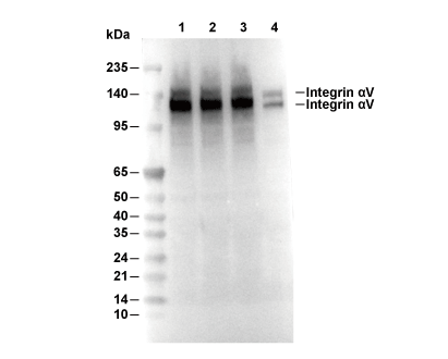 Lane 1: A549, Lane 2: MCF7, Lane 3: Jurkat, Lane 4: RL-7
Fundamentos del experimento
WB
Recommended SDS-PAGE separating gel concentration: 5%.
Recommended SDS-PAGE separating gel concentration: 5%.
Información de uso
| Dilución |
|---|
|
| Aplicación |
|---|
| WB, IF |
| Reactividad |
|---|
| Human, Mouse, Rat, Monkey |
| Fuente |
|---|
| Rabbit Monoclonal Antibody |
| Tampón de almacenamiento |
|---|
| PBS, pH 7.2+50% Glycerol+0.05% BSA+0.01% NaN3 |
| Almacenamiento (desde la fecha de recepción) |
|---|
| -20°C (avoid freeze-thaw cycles), 2 years |
| PM predicho |
|---|
| 140 kDa, 135 kDa |
| Control positivo | A2780 cells; A549 cells; MCF7cells; Jurkat cells; RL-7cells |
|---|---|
| Control negativo |
Métodos experimentales
| WB |
|---|
Experimental Protocol:
Sample preparation
1. Tissue: Lyse the tissue sample by adding an appropriate volume of ice-cold RIPA/NP-40 Lysis Buffer (containing Protease Inhibitor Cocktail),and homogenize the tissue at a low temperature. 2. Adherent cell: Aspirate the culture medium and wash the cells with ice-cold PBS twice. Lyse the cells by adding an appropriate volume of RIPA/NP-40 Lysis Buffer (containing Protease Inhibitor Cocktail) and put the sample on ice for 5 min. 3. Suspension cell: Transfer the culture medium to a pre-cooled centrifuge tube. Centrifuge and aspirate the supernatant. Wash the cells with ice-cold PBS twice. Lyse the cells by adding an appropriate volume of RIPA/NP-40 Lysis Buffer (containing Protease Inhibitor Cocktail) and put the sample on ice for 5 min. 4. Place the lysate into a pre-cooled microcentrifuge tube. Centrifuge at 4°C for 15 min. Collect the supernatant;
5. Remove a small volume of lysate to determine the protein concentration;
6. Combine the lysate with protein loading buffer. Boil 20 µL sample under 95-100°C for 5 min. Centrifuge for 5 min after cool down on ice.
Electrophoretic separation
1. According to the concentration of extracted protein, load appropriate amount of protein sample and marker onto SDS-PAGE gels for electrophoresis. Recommended separating gel (lower gel) concentration: 5%. Reference Table for Selecting SDS-PAGE Separation Gel Concentrations 2. Power up 80V for 30 minutes. Then the power supply is adjusted (110 V~150 V), the Marker is observed, and the electrophoresis can be stopped when the indicator band of the predyed protein Marker where the protein is located is properly separated. (Note that the current should not be too large when electrophoresis, too large current (more than 150 mA) will cause the temperature to rise, affecting the result of running glue. If high currents cannot be avoided, an ice bath can be used to cool the bath.)
Transfer membrane
1. Take out the converter, soak the clip and consumables in the pre-cooled converter;
2. Activate PVDF membrane with methanol for 1 min and rinse with transfer buffer;
3. Install it in the order of "black edge of clip - sponge - filter paper - filter paper - glue -PVDF membrane - filter paper - filter paper - sponge - white edge of clip"; 4. The protein was electrotransferred to PVDF membrane. ( 0.45 µm PVDF membrane is recommended ) Reference Table for Selecting PVDF Membrane Pore Size Specifications Recommended conditions for wet transfer: 200 mA, 120 min. ( Note that the transfer conditions can be adjusted according to the protein size. For high-molecular-weight proteins, a higher current and longer transfer time are recommended. However, ensure that the transfer tank remains at a low temperature to prevent gel melting.)
Block
1. After electrotransfer, wash the film with TBST at room temperature for 5 minutes;
2. Incubate the film in the blocking solution for 1 hour at room temperature;
3. Wash the film with TBST for 3 times, 5 minutes each time.
Antibody incubation
1. Use 5% skim milk powder to prepare the primary antibody working liquid (recommended dilution ratio for primary antibody 1:1000), gently shake and incubate with the film at 4°C overnight; 2. Wash the film with TBST 3 times, 5 minutes each time;
3. Add the secondary antibody to the blocking solution and incubate with the film gently at room temperature for 1 hour;
4. After incubation, wash the film with TBST 3 times for 5 minutes each time.
Antibody staining
1. Add the prepared ECL luminescent substrate (or select other color developing substrate according to the second antibody) and mix evenly;
2. Incubate with the film for 1 minute, remove excess substrate (keep the film moist), wrap with plastic film, and expose in the imaging system. |
| IF |
|---|
Experimental Protocol:
Sample Preparation
1. Adherent Cells: Place a clean, sterile coverslip in a culture dish. Once the cells grow to near confluence as a monolayer, remove the coverslip for further use.
2. Suspension Cells: Seed the cells onto a clean, sterile slide coated with poly-L-lysine.
3. Frozen Sections: Allow the slide to thaw at room temperature. Wash it with pure water or PBS for 2 times, 3 minutes each time.
4. Paraffin Sections: Deparaffinization and rehydration. Wash the slide with pure water or PBS for 3 times, 3 minutes each time. Then perform antigen retrieval.
Fixation
1. Fix the cell coverslips/spots or tissue sections at room temperature using a fixative such as 4% paraformaldehyde (4% PFA) for 10-15 minutes.
2. Wash the sample with PBS for 3 times, 3 minutes each time.
Permeabilization
1.Add a detergent such as 0.1–0.3% Triton X-100 to the sample and incubate at room temperature for 10–20 minutes.
(Note: This step is only required for intracellular antigens. For antigens expressed on the cell membrane, this step is unnecessary.)
Wash the sample with PBS for 3 times, 3 minutes each time.
Blocking
Add blocking solution and incubate at room temperature for at least 1 hour. (Common blocking solutions include: serum from the same source as the secondary antibody, BSA, or goat serum.)
Note: Ensure the sample remains moist during and after the blocking step to prevent drying, which can lead to high background.
Immunofluorescence Staining (Day 1)
1. Remove the blocking solution and add the diluted primary antibody.
2. Incubate the sample in a humidified chamber at 4°C overnight.
Immunofluorescence Staining (Day 2)
1. Remove the primary antibody and wash with PBST for 3 times, 5 minutes each time.
2. Add the diluted fluorescent secondary antibody and incubate in the dark at 4°C for 1–2 hours.
3. Remove the secondary antibody and wash with PBST for 3 times, 5 minutes each time.
4. Add diluted DAPI and incubate at room temperature in the dark for 5–10 minutes.
5. Wash with PBST for 3 times, 5 minutes each time.
Mounting
1. Mount the sample with an anti-fade mounting medium.
2. Allow the slide to dry at room temperature overnight in the dark.
3. Store the slide in a slide storage box at 4°C, protected from light.
|
Descripción biológica
| Especificidad |
|---|
| Integrin αV Antibody [P12L6] detects endogenous levels of total Integrin αV protein. |
| Localización subcelular |
|---|
| Cell junction, Cell membrane, Membrane |
| Uniprot ID |
|---|
| P06756 |
| Clon |
|---|
| P12L6 |
| Sinónimo |
|---|
| Integrin alpha-V; Vitronectin receptor; Vitronectin receptor subunit alpha; Integrin alpha-V heavy chain; Integrin alpha-V light chain; CD51 |
| Antecedentes |
|---|
| Integrin αV, encoded by the ITGAV gene, is a versatile integrin alpha subunit that forms heterodimers with β1, β3, β5, β6, or β8 subunits, mediating cell-extracellular matrix (ECM) adhesion by recognizing RGD motifs in ligands such as vitronectin, fibronectin, and osteopontin, and playing key roles in cell migration, signaling, and tissue remodeling. The type I transmembrane glycoprotein features a large extracellular domain with a seven-bladed β-propeller head (lacking an I-domain), thigh, and two calf domains organized in a bent, low-affinity resting conformation that transitions to an extended, high-affinity state upon inside-out activation via talin/kindlin binding to its short cytoplasmic tail. The ligand-binding pocket is formed at the αV propeller-βI domain interface and is stabilized by metal ion sites (MIDAS, ADMIDAS, SyMBS on β; Ca²⁺ sites on αV genu/propeller). αV integrins regulate proliferation, survival, and migration through outside-in signaling via FAK/Src, PI3K/Akt, and MAPK/ERK pathways, with specific heterodimers mediating angiogenesis (αVβ3/β5 in endothelium), TGF-β activation (αVβ6/β8 in fibrosis and immunity), and osteoclast-mediated bone resorption (αVβ3). Dysregulation of αV integrins drives tumor progression, metastasis, and therapeutic resistance in cancers (pancreatic, glioblastoma, breast), contributes to pulmonary fibrosis and osteoporosis, and facilitates viral entry (adenovirus, SARS-CoV-2), establishing αV as a validated therapeutic target for antibodies and RGD mimetics in oncology and antifibrotic treatments. |
| Referencias |
|---|
|
Soporte técnico
Tel: +1-832-582-8158 Ext:3
Si tiene alguna otra consulta, por favor deje un mensaje.
Los productos son solo para uso de investigación. No para uso humano. No vendemos a pacientes.
©Copyright 2013 Selleck Chemicals. Todos los derechos reservados.