Solo para uso en investigación
IKKα + IKKβ Antibody [H21N10]
Nº de cat.: F1703
-
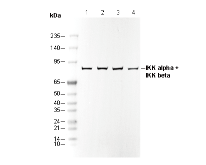 Lane 1: A431
Lane 2: 293T
Lane 3: RAW 264.7
Lane 4: NIH/3T3
Información de uso
| Dilución |
|---|
|
| Aplicación |
|---|
| WB, IP |
| Reactividad |
|---|
| Human, Mouse, Rat |
| Fuente |
|---|
| Rabbit Monoclonal Antibody |
| Tampón de almacenamiento |
|---|
| PBS, pH 7.2+50% Glycerol+0.05% BSA+0.01% NaN₃ |
| Almacenamiento (desde la fecha de recepción) |
|---|
| –20°C (avoid freeze-thaw cycles), 2 years |
| PM predicho PM observado |
|---|
| 87 kDa 75 kDa, 87 kDa |
| *¿Por qué difieren los pesos moleculares predicho y real? Las siguientes razones pueden explicar las diferencias entre el peso molecular de la proteína predicho y real. |
| Control positivo | Human fetal kidney; Mouse brain; Mouse kidney; Mouse spleen; Rat kidney; HeLa; Daudi; HL-60; A431; 293T; C6; Raw264.7; NIH/3T3; Jurkat |
|---|---|
| Control negativo |
Métodos experimentales
| WB |
|---|
Experimental Protocol:
Sample preparation
1. Tissue: Lyse the tissue sample by adding an appropriate volume of ice-cold RIPA/NP-40 Lysis Buffer (containing Protease Inhibitor Cocktail),and homogenize the tissue at a low temperature or lyse it by sonication on ice, then incubate on ice for 30 minutes. 2. Adherent cell: Aspirate the culture medium and transfer the cells into an EP tube. Wash the cells with ice-cold PBS twice. Add an appropriate volume of RIPA/NP-40 Lysis Buffer (containing Protease Inhibitor Cocktail), sonicate to lyse the cells, and incubate on ice for 30 minutes. 3. Suspension cell: Transfer the culture medium to a pre-cooled centrifuge tube. Centrifuge and aspirate the supernatant. Wash the cells with ice-cold PBS twice.Add an appropriate volume of RIPA/NP-40 Lysis Buffer (containing Protease Inhibitor Cocktail), sonicate to lyse the cells, and incubate on ice for 30 minutes. 4. Place the lysate into a pre-cooled microcentrifuge tube. Centrifuge at 4°C for 15 min. Collect the supernatant;
5. Remove a small volume of lysate to determine the protein concentration;
6. Combine the lysate with protein loading buffer. Boil 20 µL sample under 95-100°C for 5 min. Centrifuge for 5 min after cool down on ice.
Electrophoretic separation
1. According to the concentration of extracted protein, load appropriate amount of protein sample and marker onto SDS-PAGE gels for electrophoresis. Recommended separating gel (lower gel) concentration: 10%. Reference Table for Selecting SDS-PAGE Separation Gel Concentrations 2. Power up 80V for 30 minutes. Then the power supply is adjusted (110 V~150 V), the Marker is observed, and the electrophoresis can be stopped when the indicator band of the predyed protein Marker where the protein is located is properly separated. (Note that the current should not be too large when electrophoresis, too large current (more than 150 mA) will cause the temperature to rise, affecting the result of running glue. If high currents cannot be avoided, an ice bath can be used to cool the bath.)
Transfer membrane
1. Take out the converter, soak the clip and consumables in the pre-cooled converter;
2. Activate PVDF membrane with methanol for 1 min and rinse with transfer buffer;
3. Install it in the order of "black edge of clip - sponge - filter paper - filter paper - glue -PVDF membrane - filter paper - filter paper - sponge - white edge of clip"; 4. The protein was electrotransferred to PVDF membrane. ( 0.45 µm PVDF membrane is recommended ) Reference Table for Selecting PVDF Membrane Pore Size Specifications Recommended conditions for wet transfer: 200 mA, 120 min. ( Note that the transfer conditions can be adjusted according to the protein size. For high-molecular-weight proteins, a higher current and longer transfer time are recommended. However, ensure that the transfer tank remains at a low temperature to prevent gel melting.)
Block
1. After electrotransfer, wash the film with TBST at room temperature for 5 minutes;
2. Incubate the film in the blocking solution for 1 hour at room temperature;
3. Wash the film with TBST for 3 times, 5 minutes each time.
Antibody incubation
1. Use 5% skim milk powder to prepare the primary antibody working liquid (recommended dilution ratio for primary antibody 1:1000), gently shake and incubate with the film at 4°C overnight; 2. Wash the film with TBST 3 times, 5 minutes each time;
3. Add the secondary antibody to the blocking solution and incubate with the film gently at room temperature for 1 hour;
4. After incubation, wash the film with TBST 3 times for 5 minutes each time.
Antibody staining
964. Add the prepared ECL luminescent substrate (or select other color developing substrate according to the second antibody) and mix evenly;
2. Incubate with the film for 1 minute, remove excess substrate (keep the film moist), wrap with plastic film, and expose in the imaging system. |
Descripción biológica
| Especificidad |
|---|
IKKα + IKKβ Antibody [H21N10] recognizes endogenous levels of total IKK α/β protein. |
| Localización subcelular |
|---|
| Cytoplasm, Membrane, Nucleus |
| Uniprot ID |
|---|
| O14920, O15111 |
| Clon |
|---|
| H21N10 |
| Antecedentes |
|---|
The NF-κB/Rel transcription factors are typically found in the cytoplasm in an inactive state, bound to inhibitory IκB proteins. Activation of NF-κB is commonly triggered through a pathway that involves the phosphorylation-induced degradation of IκB via the proteasome. A crucial step in this process is the activation of the IκB kinase (IKK) complex, a high molecular weight assembly comprising three closely associated subunits. IKKα and IKKβ function as the kinase's catalytic subunits, while IKKγ acts as the regulatory component. For IKK activation, phosphorylation occurs at Ser177 and Ser181 in the activation loop of IKKβ (corresponding to Ser176 and Ser180 in IKKα), leading to conformational changes that activate the kinase. |
| Referencias |
|---|
|
Soporte técnico
Tel: +1-832-582-8158 Ext:3
Si tiene alguna otra consulta, por favor deje un mensaje.
Los productos son solo para uso de investigación. No para uso humano. No vendemos a pacientes.
©Copyright 2013 Selleck Chemicals. Todos los derechos reservados.