|
Cómo citar 1. Para citas en el texto (Materiales y métodos): 2. Para la tabla de recursos clave: |
||
|
Llamada gratuita: (877) 796-6397 -- Solo EE. UU. y Canadá -- |
Fax: +1-832-582-8590 Pedidos: +1-832-582-8158 |
Soporte técnico: +1-832-582-8158 Ext:3 Por favor, indique su número de pedido en el correo electrónico. Nos esforzamos por responder a todas las consultas por correo electrónico en el plazo de un día hábil. |
Descripción biológica
| Especificidad | Topoisomerase IIα Antibody [E2A15] detecta niveles endógenos de la proteína total Topoisomerase II α. |
|---|---|
| Antecedentes | Las topoisomerasas de tipo IIA (Top2) son enzimas fundamentales que regulan la topología del ADN y la organización cromosómica en varios organismos, cruciales para la replicación del ADN, la transcripción y la segregación cromosómica. En humanos, existen dos parálogos, la topoisomerasa IIα (TOP2A) y la topoisomerasa IIβ (TOP2B), que comparten una homología de secuencia significativa pero tienen patrones de expresión y funciones distintos. TOP2A se encarga principalmente de la segregación cromosómica y la replicación del ADN, mientras que TOP2B regula la transcripción. La Topoisomerase II alpha (TOP2A) está codificada por el gen TOP2A y se localiza en el cromosoma 17q12-q21. Actúa como una enzima nuclear crucial que controla los estados topológicos del ADN rompiendo transitoriamente el ADN de doble cadena, participando así en la replicación del ADN, la transcripción, la formación, el enriquecimiento y la separación de cromosomas. Sus anomalías conducen a la inestabilidad cromosómica y la tumorigenicidad, y sirven como un objetivo directo para que las antraciclinas induzcan DNA Damage. Al igual que Ki67, TOP2A también sirve como un marcador de proliferación, fuertemente expresado en células proliferativas. Su expresión es notablemente mayor en subtipos proliferativos de cáncer de mama, como las enfermedades triple negativas y enriquecidas con HER2, con una alta expresión de la proteína TOP2A asociada con un mal pronóstico. |
Información de uso
| Aplicación | WB, IHC | Dilución |
|
||||
|---|---|---|---|---|---|---|---|
| Reactividad | Human, Mouse, Rat | ||||||
| Fuente | Rabbit Monoclonal Antibody | MW | 174 kDa | ||||
| Tampón de almacenamiento | PBS, pH 7.2+50% Glycerol+0.05% BSA+0.01% NaN₃ | Almacenamiento (Desde la fecha de recepción) |
–20°C (avoid freeze-thaw cycles), 2 years | ||||
| WB |
Experimental Protocol:
Sample preparation
1. Tissue: Lyse the tissue sample by adding an appropriate volume of ice-cold RIPA/NP-40 Lysis Buffer (containing Protease Inhibitor Cocktail),and homogenize the tissue at a low temperature or lyse it by sonication on ice, then incubate on ice for 30 minutes. 2. Adherent cell: Aspirate the culture medium and transfer the cells into an EP tube. Wash the cells with ice-cold PBS twice. Add an appropriate volume of RIPA/NP-40 Lysis Buffer (containing Protease Inhibitor Cocktail), sonicate to lyse the cells, and incubate on ice for 30 minutes. 3. Suspension cell: Transfer the culture medium to a pre-cooled centrifuge tube. Centrifuge and aspirate the supernatant. Wash the cells with ice-cold PBS twice.Add an appropriate volume of RIPA/NP-40 Lysis Buffer (containing Protease Inhibitor Cocktail), sonicate to lyse the cells, and incubate on ice for 30 minutes. 4. Place the lysate into a pre-cooled microcentrifuge tube. Centrifuge at 4°C for 15 min. Collect the supernatant;
5. Remove a small volume of lysate to determine the protein concentration;
6. Combine the lysate with protein loading buffer. Boil 20 µL sample under 95-100°C for 5 min. Centrifuge for 5 min after cool down on ice.
Electrophoretic separation
1. According to the concentration of extracted protein, load appropriate amount of protein sample and marker onto SDS-PAGE gels for electrophoresis. Recommended separating gel (lower gel) concentration: 5%. Reference Table for Selecting SDS-PAGE Separation Gel Concentrations 2. Power up 80V for 30 minutes. Then the power supply is adjusted (110 V~150 V), the Marker is observed, and the electrophoresis can be stopped when the indicator band of the predyed protein Marker where the protein is located is properly separated. (Note that the current should not be too large when electrophoresis, too large current (more than 150 mA) will cause the temperature to rise, affecting the result of running glue. If high currents cannot be avoided, an ice bath can be used to cool the bath.)
Transfer membrane
1. Take out the converter, soak the clip and consumables in the pre-cooled converter;
2. Activate PVDF membrane with methanol for 1 min and rinse with transfer buffer;
3. Install it in the order of "black edge of clip - sponge - filter paper - filter paper - glue -PVDF membrane - filter paper - filter paper - sponge - white edge of clip"; 4. The protein was electrotransferred to PVDF membrane. ( 0.45 µm PVDF membrane is recommended ) Reference Table for Selecting PVDF Membrane Pore Size Specifications Recommended conditions for wet transfer: 200 mA, 120 min. ( Note that the transfer conditions can be adjusted according to the protein size. For high-molecular-weight proteins, a higher current and longer transfer time are recommended. However, ensure that the transfer tank remains at a low temperature to prevent gel melting.)
Block
1. After electrotransfer, wash the film with TBST at room temperature for 5 minutes;
2. Incubate the film in the blocking solution for 1 hour at room temperature;
3. Wash the film with TBST for 3 times, 5 minutes each time.
Antibody incubation
1. Use 5% skim milk powder to prepare the primary antibody working liquid (recommended dilution ratio for primary antibody 1:10000), gently shake and incubate with the film at 4°C overnight; 2. Wash the film with TBST 3 times, 5 minutes each time;
3. Add the secondary antibody to the blocking solution and incubate with the film gently at room temperature for 1 hour;
4. After incubation, wash the film with TBST 3 times for 5 minutes each time.
Antibody staining
566. Add the prepared ECL luminescent substrate (or select other color developing substrate according to the second antibody) and mix evenly;
2. Incubate with the film for 1 minute, remove excess substrate (keep the film moist), wrap with plastic film, and expose in the imaging system.
|
Referencias
|
Datos de aplicación
WB
Validado por Selleck
-
 Lane 1: C2C12 (Preparation by RIPA lysis method)
Lane 2: C2C12 (Preparation by 1% SDS thermal cracking method)
Lane 3: Jurkat
Lane 4: Neuro-2a
Lane 5: Human breast cancer
Lane 6: Mouse thymus
Lane 7: Rat thymus
Lane 8: Rat testis
IHC
Validado por Selleck
-
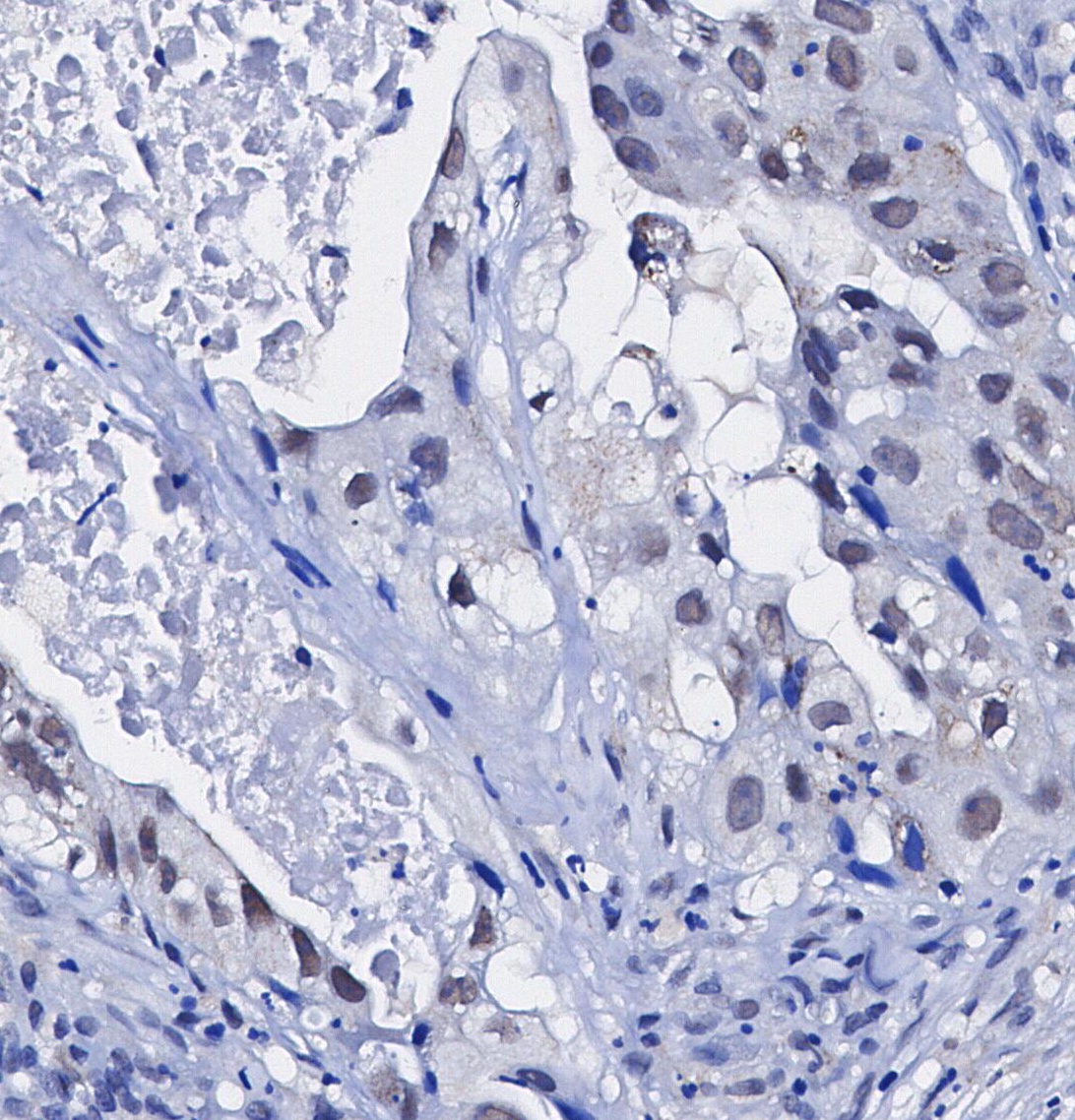 Immunohistochemical analysis of formalin fixed paraffin embedded human Lung adenocarcinoma tissue with F1219 at 1/100 dilution.