|
Cómo citar 1. Para citas en el texto (Materiales y métodos): 2. Para la tabla de recursos clave: |
||
|
Llamada gratuita: (877) 796-6397 -- Solo EE. UU. y Canadá -- |
Fax: +1-832-582-8590 Pedidos: +1-832-582-8158 |
Soporte técnico: +1-832-582-8158 Ext:3 Por favor, indique su número de pedido en el correo electrónico. Nos esforzamos por responder a todas las consultas por correo electrónico en el plazo de un día hábil. |
Descripción biológica
| Especificidad | STAT5 Antibody [B9G7] detecta niveles endógenos de proteína Stat5 total. Este anticuerpo detecta las isoformas alfa y beta de Stat5 (Stat5a y Stat5b). |
|---|---|
| Antecedentes | El transductor de señal y activador de la transcripción 5 (STAT5) comprende dos isoformas altamente homólogas, STAT5A y STAT5B, que funcionan como mediadores clave de la señalización de citocinas y factores de crecimiento. Estos factores de transcripción son activados por las Janus quinasas (JAK1, JAK2 y JAK3) tras la unión del ligando a los receptores de citocinas. Un amplio espectro de ligandos —incluyendo interleucina-2 (IL-2), factor estimulante de colonias de granulocitos y macrófagos (GM-CSF), hormona del crecimiento y prolactina— puede desencadenar la activación de STAT5. Tras la estimulación del receptor, las proteínas STAT5 sufren fosforilación de tirosina, se dimerizan (ya sea como homo- o heterodímeros) y se translocan al núcleo, donde regulan la transcripción de los genes target. La señalización de STAT5 es fundamental para el control de diversos procesos biológicos, incluyendo la proliferación celular, la diferenciación, la supervivencia y funciones específicas de tejidos como la hematopoyesis y el metabolismo de los hepatocitos. La actividad de STAT5 debe ser regulada con precisión: la hiperactivación contribuye a estados patológicos como el cáncer, los trastornos mieloproliferativos, la inflamación crónica y las enfermedades autoinmunes, mientras que una activación insuficiente puede resultar en anemia, trombocitopenia, enanismo, infertilidad, inmunodeficiencia y disfunción metabólica. Tanto STAT5A como STAT5B existen en múltiples isoformas más cortas generadas a través de empalme alternativo y procesamiento post-traduccional. Estas proteínas son moduladas además por fosforilación (en residuos de serina, treonina o tirosina), ubiquitinación y glicosilación, lo que influye en su estabilidad, capacidad de dimerización y rendimiento transcripcional. A través de la formación de complejos diméricos distintos, STAT5 orquesta programas de expresión génica críticos para mantener la homeostasis celular normal y coordinar las respuestas fisiológicas sistémicas. |
Información de uso
| Aplicación | WB, IP, ChIP | Dilución |
|
||||||
|---|---|---|---|---|---|---|---|---|---|
| Reactividad | Mouse, Rat, Human | ||||||||
| Fuente | Rabbit Monoclonal Antibody | MW | 90 kDa | ||||||
| Tampón de almacenamiento | PBS, pH 7.2+50% Glycerol+0.05% BSA+0.01% NaN3 | Almacenamiento (Desde la fecha de recepción) |
-20°C (avoid freeze-thaw cycles), 2 years | ||||||
| WB |
Experimental Protocol:
Sample preparation
1. Tissue: Lyse the tissue sample by adding an appropriate volume of ice-cold RIPA/NP-40 Lysis Buffer (containing Protease Inhibitor Cocktail),and homogenize the tissue at a low temperature. 2. Adherent cell: Aspirate the culture medium and wash the cells with ice-cold PBS twice. Lyse the cells by adding an appropriate volume of RIPA/NP-40 Lysis Buffer (containing Protease Inhibitor Cocktail) and put the sample on ice for 5 min. 3. Suspension cell: Transfer the culture medium to a pre-cooled centrifuge tube. Centrifuge and aspirate the supernatant. Wash the cells with ice-cold PBS twice. Lyse the cells by adding an appropriate volume of RIPA/NP-40 Lysis Buffer (containing Protease Inhibitor Cocktail) and put the sample on ice for 5 min. 4. Place the lysate into a pre-cooled microcentrifuge tube. Centrifuge at 4°C for 15 min. Collect the supernatant;
5. Remove a small volume of lysate to determine the protein concentration;
6. Combine the lysate with protein loading buffer. Boil 20 µL sample under 95-100°C for 5 min. Centrifuge for 5 min after cool down on ice.
Electrophoretic separation
1. According to the concentration of extracted protein, load appropriate amount of protein sample and marker onto SDS-PAGE gels for electrophoresis. Recommended separating gel (lower gel) concentration: 10%. Reference Table for Selecting SDS-PAGE Separation Gel Concentrations 2. Power up 80V for 30 minutes. Then the power supply is adjusted (110 V~150 V), the Marker is observed, and the electrophoresis can be stopped when the indicator band of the predyed protein Marker where the protein is located is properly separated. (Note that the current should not be too large when electrophoresis, too large current (more than 150 mA) will cause the temperature to rise, affecting the result of running glue. If high currents cannot be avoided, an ice bath can be used to cool the bath.)
Transfer membrane
1. Take out the converter, soak the clip and consumables in the pre-cooled converter;
2. Activate PVDF membrane with methanol for 1 min and rinse with transfer buffer;
3. Install it in the order of "black edge of clip - sponge - filter paper - filter paper - glue -PVDF membrane - filter paper - filter paper - sponge - white edge of clip"; 4. The protein was electrotransferred to PVDF membrane. ( 0.45 µm PVDF membrane is recommended ) Reference Table for Selecting PVDF Membrane Pore Size Specifications Recommended conditions for wet transfer: 200 mA, 120 min. ( Note that the transfer conditions can be adjusted according to the protein size. For high-molecular-weight proteins, a higher current and longer transfer time are recommended. However, ensure that the transfer tank remains at a low temperature to prevent gel melting.)
Block
1. After electrotransfer, wash the film with TBST at room temperature for 5 minutes;
2. Incubate the film in the blocking solution for 1 hour at room temperature;
3. Wash the film with TBST for 3 times, 5 minutes each time.
Antibody incubation
1. Use 5% skim milk powder to prepare the primary antibody working liquid (recommended dilution ratio for primary antibody 1:1000), gently shake and incubate with the film at 4°C overnight; 2. Wash the film with TBST 3 times, 5 minutes each time;
3. Add the secondary antibody to the blocking solution and incubate with the film gently at room temperature for 1 hour;
4. After incubation, wash the film with TBST 3 times for 5 minutes each time.
Antibody staining
1. Add the prepared ECL luminescent substrate (or select other color developing substrate according to the second antibody) and mix evenly;
2. Incubate with the film for 1 minute, remove excess substrate (keep the film moist), wrap with plastic film, and expose in the imaging system.
|
Referencias
|
Datos de aplicación
WB
Validado por Selleck
-
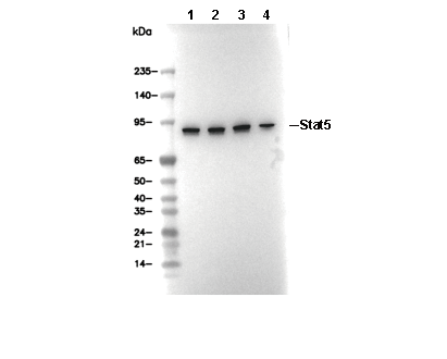 Lane 1: K562, Lane 2: MOLT4, Lane 3: A20, Lane 4: Daudi