|
Cómo citar 1. Para citas en el texto (Materiales y métodos): 2. Para la tabla de recursos clave: |
||
|
Llamada gratuita: (877) 796-6397 -- Solo EE. UU. y Canadá -- |
Fax: +1-832-582-8590 Pedidos: +1-832-582-8158 |
Soporte técnico: +1-832-582-8158 Ext:3 Por favor, indique su número de pedido en el correo electrónico. Nos esforzamos por responder a todas las consultas por correo electrónico en el plazo de un día hábil. |
Descripción biológica
| Especificidad | SOX9 Antibody [D1B10] reconoce los niveles endógenos de la proteína Sox9 total. |
|---|---|
| Antecedentes | SOX9 (SRY-box transcription factor 9) es un factor de transcripción del desarrollo clave, miembro de la familia SOX (SRY-type HMG box), esencial para la organogénesis de mamíferos. Expresado en una amplia variedad de tejidos —incluyendo cartílago, testículos, sistema nervioso, corazón, pulmón, páncreas y más— SOX9 desempeña papeles críticos en la determinación del destino celular, el mantenimiento de progenitores y la regulación génica específica de tejidos. Codifica una proteína de 509 aminoácidos con dominios funcionales distintos: un dominio de unión al ADN High-mobility Group (HMG), un dominio de dimerización (DIM), dos dominios de transactivación (TAM y TAC) y un dominio rico en prolina/glutamina/alanina (PQA). Estos dominios permiten a SOX9 unirse al ADN, formar dímeros o heterodímeros, reclutar coactivadores como CBP/p300 y MED12, y regular la expresión génica de manera específica al contexto. Su expresión está estrictamente regulada por potenciadores de largo alcance específicos de tejido ubicados dentro de un gran desierto génico que rodea el locus SOX9 en el cromosoma 17q (humano) o 11q (ratón), lo que permite un control espacial y temporal preciso durante el desarrollo. Funcionalmente, SOX9 es esencial para el desarrollo de múltiples órganos, incluidos huesos, testículos, corazón, pulmón, páncreas, intestino y sistema nervioso. Las mutaciones en el gen SOX9 humano provocaron displasia campomélica, un trastorno de haploinsuficiencia con varias malformaciones esqueléticas frecuentemente acompañadas de inversión sexual 46, XY. |
Información de uso
| Aplicación | WB, IP, IHC, IF, FCM | Dilución |
|
||||||||||
|---|---|---|---|---|---|---|---|---|---|---|---|---|---|
| Reactividad | Human, Mouse, Rat | ||||||||||||
| Fuente | Rabbit Monoclonal Antibody | MW | 56 kDa | ||||||||||
| Tampón de almacenamiento | PBS, pH 7.2+50% Glycerol+0.05% BSA+0.01% NaN3 | Almacenamiento (Desde la fecha de recepción) |
-20°C (avoid freeze-thaw cycles), 2 years | ||||||||||
| WB |
Experimental Protocol:
Sample preparation
1. Tissue: Lyse the tissue sample by adding an appropriate volume of ice-cold RIPA/NP-40 Lysis Buffer (containing Protease Inhibitor Cocktail),and homogenize the tissue at a low temperature or lyse it by sonication on ice, then incubate on ice for 30 minutes. 2. Adherent cell: Aspirate the culture medium and transfer the cells into an EP tube. Wash the cells with ice-cold PBS twice. Add an appropriate volume of RIPA/NP-40 Lysis Buffer (containing Protease Inhibitor Cocktail), sonicate to lyse the cells, and incubate on ice for 30 minutes. 3. Suspension cell: Transfer the culture medium to a pre-cooled centrifuge tube. Centrifuge and aspirate the supernatant. Wash the cells with ice-cold PBS twice.Add an appropriate volume of RIPA/NP-40 Lysis Buffer (containing Protease Inhibitor Cocktail), sonicate to lyse the cells, and incubate on ice for 30 minutes. 4. Place the lysate into a pre-cooled microcentrifuge tube. Centrifuge at 4°C for 15 min. Collect the supernatant;
5. Remove a small volume of lysate to determine the protein concentration;
6. Combine the lysate with protein loading buffer. Boil 20 µL sample under 95-100°C for 5 min. Centrifuge for 5 min after cool down on ice.
Electrophoretic separation
1. According to the concentration of extracted protein, load appropriate amount of protein sample and marker onto SDS-PAGE gels for electrophoresis. Recommended separating gel (lower gel) concentration: 10%. Reference Table for Selecting SDS-PAGE Separation Gel Concentrations 2. Power up 80V for 30 minutes. Then the power supply is adjusted (110 V~150 V), the Marker is observed, and the electrophoresis can be stopped when the indicator band of the predyed protein Marker where the protein is located is properly separated. (Note that the current should not be too large when electrophoresis, too large current (more than 150 mA) will cause the temperature to rise, affecting the result of running glue. If high currents cannot be avoided, an ice bath can be used to cool the bath.)
Transfer membrane
1. Take out the converter, soak the clip and consumables in the pre-cooled converter;
2. Activate PVDF membrane with methanol for 1 min and rinse with transfer buffer;
3. Install it in the order of "black edge of clip - sponge - filter paper - filter paper - glue -PVDF membrane - filter paper - filter paper - sponge - white edge of clip"; 4. The protein was electrotransferred to PVDF membrane. ( 0.45 µm PVDF membrane is recommended ) Reference Table for Selecting PVDF Membrane Pore Size Specifications Recommended conditions for wet transfer: 200 mA, 120 min. ( Note that the transfer conditions can be adjusted according to the protein size. For high-molecular-weight proteins, a higher current and longer transfer time are recommended. However, ensure that the transfer tank remains at a low temperature to prevent gel melting.)
Block
1. After electrotransfer, wash the film with TBST at room temperature for 5 minutes;
2. Incubate the film in the blocking solution for 1 hour at room temperature;
3. Wash the film with TBST for 3 times, 5 minutes each time.
Antibody incubation
1. Use 5% skim milk powder to prepare the primary antibody working liquid (recommended dilution ratio for primary antibody 1:1000), gently shake and incubate with the film at 4°C overnight; 2. Wash the film with TBST 3 times, 5 minutes each time;
3. Add the secondary antibody to the blocking solution and incubate with the film gently at room temperature for 1 hour;
4. After incubation, wash the film with TBST 3 times for 5 minutes each time.
Antibody staining
1389. Add the prepared ECL luminescent substrate (or select other color developing substrate according to the second antibody) and mix evenly;
2. Incubate with the film for 1 minute, remove excess substrate (keep the film moist), wrap with plastic film, and expose in the imaging system.
|
Referencias
|
Datos de aplicación
WB
Validado por Selleck
-
 Lane 1: SW480, Lane 2: HeLa, Lane 3: NIH/3T3
IF
Validado por Selleck
-
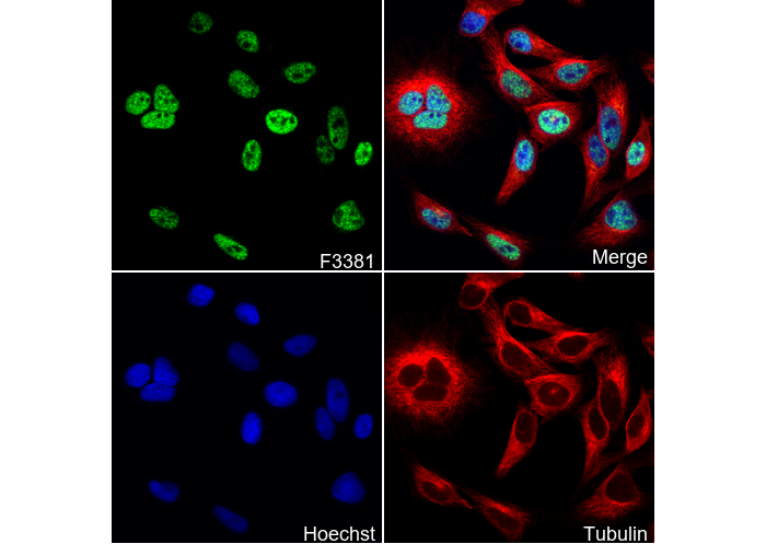 Immunofluorescent analysis of SW480 cells using F3381 (green, 1:250), Hoechst (blue) and tubulin (Red).