|
Cómo citar 1. Para citas en el texto (Materiales y métodos): 2. Para la tabla de recursos clave: |
||
|
Llamada gratuita: (877) 796-6397 -- Solo EE. UU. y Canadá -- |
Fax: +1-832-582-8590 Pedidos: +1-832-582-8158 |
Soporte técnico: +1-832-582-8158 Ext:3 Por favor, indique su número de pedido en el correo electrónico. Nos esforzamos por responder a todas las consultas por correo electrónico en el plazo de un día hábil. |
Descripción biológica
| Especificidad | SFRP4 Antibody [K23D16] reconoce los niveles endógenos de la proteína SFRP4 total. |
|---|---|
| Antecedentes | La proteína 4 relacionada con Frizzled secretada (sFRP4) es una glicoproteína soluble que desempeña un papel fundamental en la modulación de la vía de señalización Wnt, que es crucial para la proliferación, diferenciación y apoptosis celular. Estructuralmente, la sFRP4 consta de un dominio rico en cisteína (CRD) responsable de la unión a las proteínas Wnt y un dominio C-terminal (CTD) que estabiliza su estructura y mejora su afinidad de unión a los ligandos Wnt. Este CRD permite a la sFRP4 antagonizar la señalización Wnt al evitar que las proteínas Wnt interactúen con los receptores Frizzled en las superficies celulares, inhibiendo así la activación aguas abajo de vías como Wnt/β-catenina. Si bien su función principal es actuar como antagonista de la señalización Wnt, la función de la sFRP4 puede depender del contexto, ya sea inhibiendo o potenciando la actividad Wnt. Está implicada en múltiples trastornos como el cáncer, donde suprime el crecimiento tumoral, y en enfermedades metabólicas como la diabetes tipo 2, donde su expresión interrumpe la sensibilidad a la insulina. También regula la apoptosis, el desarrollo de tejidos y la homeostasis celular. También está implicada en trastornos metabólicos como la diabetes tipo 2, donde desempeña un papel en la reducción de la sensibilidad a la insulina, así como en afecciones como la psoriasis, la fibrosis renal y las enfermedades relacionadas con los huesos. |
Información de uso
| Aplicación | IHC, IF, FCM | Dilución |
|
||||||
|---|---|---|---|---|---|---|---|---|---|
| Reactividad | Human, Mouse | ||||||||
| Fuente | Rabbit Monoclonal Antibody | MW | |||||||
| Tampón de almacenamiento | PBS, pH 7.2+50% Glycerol+0.05% BSA+0.01% NaN₃ | Almacenamiento (Desde la fecha de recepción) |
-20°C (avoid freeze-thaw cycles), 2 years | ||||||
| IF |
Experimental Protocol:
Sample Preparation
1. Adherent Cells: Place a clean, sterile coverslip in a culture dish. Once the cells grow to near confluence as a monolayer, remove the coverslip for further use.
2. Suspension Cells: Seed the cells onto a clean, sterile slide coated with poly-L-lysine.
3. Frozen Sections: Allow the slide to thaw at room temperature. Wash it with pure water or PBS for 2 times, 3 minutes each time.
4. Paraffin Sections: Deparaffinization and rehydration. Wash the slide with pure water or PBS for 3 times, 3 minutes each time. Then perform antigen retrieval.
Fixation
1. Fix the cell coverslips/spots or tissue sections at room temperature using a fixative such as 4% paraformaldehyde (4% PFA) for 10-15 minutes.
2. Wash the sample with PBS for 3 times, 3 minutes each time.
Permeabilization
1.Add a detergent such as 0.1–0.3% Triton X-100 to the sample and incubate at room temperature for 10–20 minutes.
(Note: This step is only required for intracellular antigens. For antigens expressed on the cell membrane, this step is unnecessary.)
Wash the sample with PBS for 3 times, 3 minutes each time.
Blocking
Add blocking solution and incubate at room temperature for at least 1 hour. (Common blocking solutions include: serum from the same source as the secondary antibody, BSA, or goat serum.)
Note: Ensure the sample remains moist during and after the blocking step to prevent drying, which can lead to high background.
Immunofluorescence Staining (Day 1)
1. Remove the blocking solution and add the diluted primary antibody.
2. Incubate the sample in a humidified chamber at 4°C overnight.
Immunofluorescence Staining (Day 2)
1. Remove the primary antibody and wash with PBST for 3 times, 5 minutes each time.
2. Add the diluted fluorescent secondary antibody and incubate in the dark at 4°C for 1–2 hours.
3. Remove the secondary antibody and wash with PBST for 3 times, 5 minutes each time.
4. Add diluted DAPI and incubate at room temperature in the dark for 5–10 minutes.
5. Wash with PBST for 3 times, 5 minutes each time.
Mounting
1. Mount the sample with an anti-fade mounting medium.
2. Allow the slide to dry at room temperature overnight in the dark.
3. Store the slide in a slide storage box at 4°C, protected from light.
|
| IHC |
Experimental Protocol:
Deparaffinization/Rehydration
1. Deparaffinize/hydrate sections:
2. Incubate sections in three washes of xylene for 5 min each.
3. Incubate sections in two washes of 100% ethanol for 10 min each.
4. Incubate sections in two washes of 95% ethanol for 10 min each.
5. Wash sections two times in dH2O for 5 min each.
6.Antigen retrieval: For Citrate: Heat slides in a microwave submersed in 1X citrate unmasking solution until boiling is initiated; continue with 10 min at a sub-boiling temperature (95°-98°C). Cool slides on bench top for 30 min.
Staining
1. Wash sections in dH2O three times for 5 min each.
2. Incubate sections in 3% hydrogen peroxide for 10 min.
3. Wash sections in dH2O two times for 5 min each.
4. Wash sections in wash buffer for 5 min.
5. Block each section with 100–400 µl of blocking solution for 1 hr at room temperature.
6. Remove blocking solution and add 100–400 µl primary antibody diluent in to each section. Incubate overnight at 4°C.
7. Remove antibody solution and wash sections with wash buffer three times for 5 min each.
8. Cover section with 1–3 drops HRPas needed. Incubate in a humidified chamber for 30 min at room temperature.
9. Wash sections three times with wash buffer for 5 min each.
10. Add DAB Chromogen Concentrate to DAB Diluent and mix well before use.
11. Apply 100–400 µl DAB to each section and monitor closely. 1–10 min generally provides an acceptable staining intensity.
12. Immerse slides in dH2O.
13. If desired, counterstain sections with hematoxylin.
14. Wash sections in dH2O two times for 5 min each.
15. Dehydrate sections: Incubate sections in 95% ethanol two times for 10 sec each; Repeat in 100% ethanol, incubating sections two times for 10 sec each; Repeat in xylene, incubating sections two times for 10 sec each.
16. Mount sections with coverslips and mounting medium.
|
Referencias
|
Datos de aplicación
IHC
Validado por Selleck
-
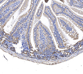 Immunohistochemical analysis of formalin fixed paraffin embedded mouse intestine tissue with F2563 at 1:50 dilution.
IF
Validado por Selleck
-
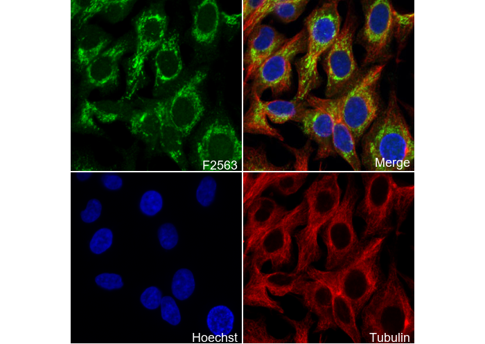 Immunofluorescent analysis of Raw264.7 cells using F2563 (green, 1:200), Hoechst (blue) and tubulin (Red).