|
Cómo citar 1. Para citas en el texto (Materiales y métodos): 2. Para la tabla de recursos clave: |
||
|
Llamada gratuita: (877) 796-6397 -- Solo EE. UU. y Canadá -- |
Fax: +1-832-582-8590 Pedidos: +1-832-582-8158 |
Soporte técnico: +1-832-582-8158 Ext:3 Por favor, indique su número de pedido en el correo electrónico. Nos esforzamos por responder a todas las consultas por correo electrónico en el plazo de un día hábil. |
Descripción biológica
| Especificidad | Phospho-Synapsin-1 (Ser605) Antibody [M6M7] detecta niveles endógenos de la proteína Synapsin-1 solo cuando está fosforilada en Ser605 (corresponde a Ser603 en rata). |
|---|---|
| Antecedentes | La Sinapsina I (proteína I) es una fosfoproteína importante específica de neuronas y un sustrato endógeno clave para las proteínas quinasas dependientes de cAMP y Ca²⁺/calmodulina. Se distribuye ampliamente en las sinapsis de todo el sistema nervioso central y periférico, donde se asocia específicamente con la superficie citoplasmática de las membranas de las vesículas sinápticas. La familia de proteínas sinapsina consta de cuatro miembros homólogos: las sinapsinas Ia e Ib (denominadas colectivamente sinapsina I) y las sinapsinas IIa y IIb (denominadas colectivamente sinapsina II). Juntas, las sinapsinas I y II representan aproximadamente el 9% del total de proteínas de las vesículas sinápticas. Las sinapsinas I y II están presentes principalmente en sinapsis maduras, mientras que la sinapsina III se expresa principalmente durante el desarrollo sináptico y en niveles comparativamente más bajos. Funcionalmente, la sinapsina I desempeña un papel crítico en la regulación de la liberación de neurotransmisores. En las neuronas, ayuda a controlar la disponibilidad de vesículas sinápticas para la exocitosis. La fosforilación en el residuo Ser-9 (fosfo-Ser9 sinapsina I) hace que la proteína se disocie de las vesículas sinápticas, un proceso esencial para la liberación de neurotransmisores. Además, la sinapsina I contribuye a la plasticidad sináptica al influir tanto en la liberación de vesículas pre- como postsinápticas. Genéticamente, las mutaciones en el gen de la sinapsina I se han relacionado con la epilepsia ligada al cromosoma X con discapacidades de aprendizaje variables y trastornos del comportamiento (XELBD), una afección neurológica caracterizada por combinaciones variables de epilepsia, deterioro cognitivo, macrocefalia y comportamiento agresivo. Ser605 se confirma como un sitio de fosforilación importante en la Sinapsina I, con evidencia in vivo que respalda su importancia. La fosforilación en Ser605 (junto con Ser568) por la proteína quinasa II dependiente de Ca²⁺/calmodulina (CaMKII) altera la capacidad de la Sinapsina I para agrupar filamentos de actina. Este es probablemente un mecanismo que regula la organización dinámica del citoesqueleto presináptico. |
Información de uso
| Aplicación | WB | Dilución |
|
||
|---|---|---|---|---|---|
| Reactividad | Human, Mouse, Rat | ||||
| Fuente | Rabbit Monoclonal Antibody | MW | 75-90 kDa | ||
| Tampón de almacenamiento | PBS, pH 7.2+50% Glycerol+0.05% BSA+0.01% NaN3 | Almacenamiento (Desde la fecha de recepción) |
-20°C (avoid freeze-thaw cycles), 2 years | ||
| WB |
Experimental Protocol:
Sample preparation
1. Tissue: Lyse the tissue sample by adding an appropriate volume of ice-cold RIPA/NP-40 Lysis Buffer (containing Protease Inhibitor Cocktail, Phosphatase Inhibitor Cocktail),and homogenize the tissue at a low temperature. 2. Adherent cell: Aspirate the culture medium and wash the cells with ice-cold PBS twice. Lyse the cells by adding an appropriate volume of RIPA/NP-40 Lysis Buffer (containing Protease Inhibitor Cocktail, Phosphatase Inhibitor Cocktail) and put the sample on ice for 5 min. 3. Suspension cell: Transfer the culture medium to a pre-cooled centrifuge tube. Centrifuge and aspirate the supernatant. Wash the cells with ice-cold PBS twice. Lyse the cells by adding an appropriate volume of RIPA/NP-40 Lysis Buffer (containing Protease Inhibitor Cocktail, Phosphatase Inhibitor Cocktail) and put the sample on ice for 5 min. 4. Place the lysate into a pre-cooled microcentrifuge tube. Centrifuge at 4°C for 15 min. Collect the supernatant;
5. Remove a small volume of lysate to determine the protein concentration;
6. Combine the lysate with protein loading buffer. Boil 20 µL sample under 95-100°C for 5 min. Centrifuge for 5 min after cool down on ice.
Electrophoretic separation
1. According to the concentration of extracted protein, load appropriate amount of protein sample and marker onto SDS-PAGE gels for electrophoresis. Recommended separating gel (lower gel) concentration: 10%. Reference Table for Selecting SDS-PAGE Separation Gel Concentrations 2. Power up 80V for 30 minutes. Then the power supply is adjusted (110 V~150 V), the Marker is observed, and the electrophoresis can be stopped when the indicator band of the predyed protein Marker where the protein is located is properly separated. (Note that the current should not be too large when electrophoresis, too large current (more than 150 mA) will cause the temperature to rise, affecting the result of running glue. If high currents cannot be avoided, an ice bath can be used to cool the bath.)
Transfer membrane
1. Take out the converter, soak the clip and consumables in the pre-cooled converter;
2. Activate PVDF membrane with methanol for 1 min and rinse with transfer buffer;
3. Install it in the order of "black edge of clip - sponge - filter paper - filter paper - glue -PVDF membrane - filter paper - filter paper - sponge - white edge of clip"; 4. The protein was electrotransferred to PVDF membrane. ( 0.45 µm PVDF membrane is recommended ) Reference Table for Selecting PVDF Membrane Pore Size Specifications Recommended conditions for wet transfer: 200 mA, 120 min. ( Note that the transfer conditions can be adjusted according to the protein size. For high-molecular-weight proteins, a higher current and longer transfer time are recommended. However, ensure that the transfer tank remains at a low temperature to prevent gel melting.)
Block
1. After electrotransfer, wash the film with TBST at room temperature for 5 minutes;
2. Incubate the film in the blocking solution ( recommending 5% BSA solution)
for 1 hour at room temperature;
3. Wash the film with TBST for 3 times, 5 minutes each time.
Antibody incubation
1. Use 5% skim milk powder to prepare the primary antibody working liquid (recommended dilution ratio for primary antibody 1:1000), gently shake and incubate with the film at 4°C overnight; 2. Wash the film with TBST 3 times, 5 minutes each time;
3. Add the secondary antibody to the blocking solution and incubate with the film gently at room temperature for 1 hour;
4. After incubation, wash the film with TBST 3 times for 5 minutes each time.
Antibody staining
1. Add the prepared ECL luminescent substrate (or select other color developing substrate according to the second antibody) and mix evenly;
2. Incubate with the film for 1 minute, remove excess substrate (keep the film moist), wrap with plastic film, and expose in the imaging system. (Exposure time of at least 150s is recommended)
|
Referencias
|
Datos de aplicación
WB
Validado por Selleck
-
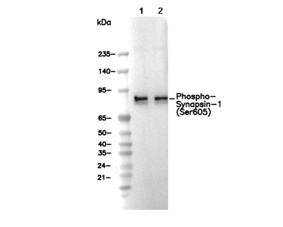 Lane 1: Mouse brain, Lane 2: Rat brain