|
Cómo citar 1. Para citas en el texto (Materiales y métodos): 2. Para la tabla de recursos clave: |
||
|
Llamada gratuita: (877) 796-6397 -- Solo EE. UU. y Canadá -- |
Fax: +1-832-582-8590 Pedidos: +1-832-582-8158 |
Soporte técnico: +1-832-582-8158 Ext:3 Por favor, indique su número de pedido en el correo electrónico. Nos esforzamos por responder a todas las consultas por correo electrónico en el plazo de un día hábil. |
Descripción biológica
| Especificidad | El anticuerpo Phospho-Rpb1 CTD (Ser2) [L19G24] reconoce los niveles endógenos de Rpb1 solo cuando la repetición del heptapéptido del dominio carboxilo-terminal (CTD) [Tyr1, Ser2, Pro3, Thr4, Ser5, Pro6, Ser7] está fosforilada en Ser2. Este anticuerpo no reacciona de forma cruzada con Rpb1 CTD fosforilado en Ser5 o Ser7. |
|---|---|
| Antecedentes | La ARN polimerasa II (RNAPII) es un complejo multiproteico responsable de transcribir el ADN en ARN, utilizando ribonucleósidos trifosfato como bloques de construcción. Su subunidad más grande, la subunidad B1 de la RNAPII (Rpb1), también conocida como subunidad A de la RNAPII (POLR2A), presenta una secuencia heptapeptídica distintiva (Tyr1,Ser2,Pro3,Thr4,Ser5,Pro6,Ser7), que se repite hasta 52 veces dentro del dominio carboxilo-terminal (CTD) de la proteína. Este heptapéptido CTD sufre diversas modificaciones postraduccionales, dictando el estado funcional del complejo polimerasa. La fosforilación del CTD durante la transcripción activa coordina la transcripción con el remodelado de la cromatina y el procesamiento del ARN, regulando el reclutamiento de enzimas modificadoras de la cromatina y proteínas de procesamiento del ARN al gen transcrito. Durante la iniciación de la transcripción, la RNAPII alberga un CTD hipofosforilado y es reclutada a los promotores génicos a través de interacciones con factores de transcripción unidos al ADN y el complejo Mediador. La liberación de la RNAPII de los promotores génicos necesita la fosforilación en Ser5 por CDK7, la subunidad catalítica del factor de transcripción IIH (TFIIH). Posteriormente, la RNAPII fosforilada en Ser2/Ser5 transcribe toda la longitud del gen hasta llegar al extremo 3', donde concluye la transcripción. La RNAPII se desengage entonces del ADN y se recicla para volver a su estado hipofosforilado mediante la acción de varias fosfatasas CTD. |
Información de uso
| Aplicación | WB, IP, ChIP | Dilución |
|
||||||
|---|---|---|---|---|---|---|---|---|---|
| Reactividad | Human, Mouse, Rat, Monkey | ||||||||
| Fuente | Rabbit Monoclonal Antibody | MW | 250 kDa | ||||||
| Tampón de almacenamiento | PBS, pH 7.2+50% Glycerol+0.05% BSA+0.01% NaN₃ | Almacenamiento (Desde la fecha de recepción) |
–20°C (avoid freeze-thaw cycles), 2 years | ||||||
| WB |
Experimental Protocol:
Sample preparation
1. Tissue: Lyse the tissue sample by adding an appropriate volume of ice-cold RIPA/NP-40 Lysis Buffer (containing Protease Inhibitor Cocktail, Phosphatase Inhibitor Cocktail),and homogenize the tissue at a low temperature or lyse it by sonication on ice, then incubate on ice for 30 minutes. 2. Adherent cell: Aspirate the culture medium and transfer the cells into an EP tube. Wash the cells with ice-cold PBS twice. Add an appropriate volume of RIPA/NP-40 Lysis Buffer (containing Protease Inhibitor Cocktail, Phosphatase Inhibitor Cocktail), sonicate to lyse the cells, and incubate on ice for 30 minutes. 3. Suspension cell: Transfer the culture medium to a pre-cooled centrifuge tube. Centrifuge and aspirate the supernatant. Wash the cells with ice-cold PBS twice.Add an appropriate volume of RIPA/NP-40 Lysis Buffer (containing Protease Inhibitor Cocktail, Phosphatase Inhibitor Cocktail), sonicate to lyse the cells, and incubate on ice for 30 minutes. 4. Place the lysate into a pre-cooled microcentrifuge tube. Centrifuge at 4°C for 15 min. Collect the supernatant;
5. Remove a small volume of lysate to determine the protein concentration;
6. Combine the lysate with protein loading buffer. Boil 20 µL sample under 95-100°C for 5 min. Centrifuge for 5 min after cool down on ice.
Electrophoretic separation
1. According to the concentration of extracted protein, load appropriate amount of protein sample and marker onto SDS-PAGE gels for electrophoresis. Recommended separating gel (lower gel) concentration: 5%. Reference Table for Selecting SDS-PAGE Separation Gel Concentrations 2. Power up 80V for 30 minutes. Then the power supply is adjusted (110 V~150 V), the Marker is observed, and the electrophoresis can be stopped when the indicator band of the predyed protein Marker where the protein is located is properly separated. (Note that the current should not be too large when electrophoresis, too large current (more than 150 mA) will cause the temperature to rise, affecting the result of running glue. If high currents cannot be avoided, an ice bath can be used to cool the bath.)
Transfer membrane
1. Take out the converter, soak the clip and consumables in the pre-cooled converter;
2. Activate PVDF membrane with methanol for 1 min and rinse with transfer buffer;
3. Install it in the order of "black edge of clip - sponge - filter paper - filter paper - glue -PVDF membrane - filter paper - filter paper - sponge - white edge of clip"; 4. The protein was electrotransferred to PVDF membrane. ( 0.45 µm PVDF membrane is recommended ) Reference Table for Selecting PVDF Membrane Pore Size Specifications Recommended conditions for wet transfer: 250 mA, 180 min. ( Note that the transfer conditions can be adjusted according to the protein size. For high-molecular-weight proteins, a higher current and longer transfer time are recommended. However, ensure that the transfer tank remains at a low temperature to prevent gel melting.)
Block
1. After electrotransfer, wash the film with TBST at room temperature for 5 minutes;
2. Incubate the film in the blocking solution ( recommending 5% BSA solution)
for 1 hour at room temperature;
3. Wash the film with TBST for 3 times, 5 minutes each time.
Antibody incubation
1. Use 5% skim milk powder to prepare the primary antibody working liquid (recommended dilution ratio for primary antibody 1:1000), gently shake and incubate with the film at 4°C overnight; 2. Wash the film with TBST 3 times, 5 minutes each time;
3. Add the secondary antibody to the blocking solution and incubate with the film gently at room temperature for 1 hour;
4. After incubation, wash the film with TBST 3 times for 5 minutes each time.
Antibody staining
313. Add the prepared ECL luminescent substrate (or select other color developing substrate according to the second antibody) and mix evenly;
2. Incubate with the film for 1 minute, remove excess substrate (keep the film moist), wrap with plastic film, and expose in the imaging system.
|
| IF |
Experimental Protocol:
Specimen Preparation
1. Aspirate liquid, then cover cells to a depth of 2–3 mm with 4% Paraformaldehyde diluted in 1X PBS.
NOTE: Paraformaldehyde is toxic, use only in a fume hood.
2. Fix cells for 15 min at room temperature.
3. Aspirate fixative, rinse three times in 1X PBS for 5 min each.
4. Proceed with Immunostaining.
Immunostaining
1. Add theblocking buffer and incubate for 60 min at RT.
2. Prepare primary antibody diluent in antibody dilution buffer as recommended .
3. Aspirate blocking solution, apply diluted primary antibody.
4. Incubate overnight at 4°C.
5. Rinse three times in 1X PBS for 5 min each.
6. Incubate specimens in fluorochrome-conjugated secondary antibody diluted in antibody dilution buffer for 1–2 hr at room temperature in the dark.
7. Rinse three times in 1X PBS for 5 min each.
8. Mount slides usingmounting medium with DAPI and cover with coverslips.
9. For best results, allow mountant to cure overnight at room temperature. For long-term storage, store slides flat at 4°C protected from light.
|
| IHC |
Experimental Protocol:
Deparaffinization/Rehydration
1. Deparaffinize/hydrate sections:
2. Incubate sections in three washes of xylene for 5 min each.
3. Incubate sections in two washes of 100% ethanol for 10 min each.
4. Incubate sections in two washes of 95% ethanol for 10 min each.
5. Wash sections two times in dH2O for 5 min each.
6.Antigen retrieval: For Citrate: Heat slides in a microwave submersed in 1X citrate unmasking solution until boiling is initiated; continue with 10 min at a sub-boiling temperature (95°-98°C). Cool slides on bench top for 30 min.
Staining
1. Wash sections in dH2O three times for 5 min each.
2. Incubate sections in 3% hydrogen peroxide for 10 min.
3. Wash sections in dH2O two times for 5 min each.
4. Wash sections in wash buffer for 5 min.
5. Block each section with 100–400 µl of blocking solution for 1 hr at room temperature.
6. Remove blocking solution and add 100–400 µl primary antibody diluent in to each section. Incubate overnight at 4°C.
7. Remove antibody solution and wash sections with wash buffer three times for 5 min each.
8. Cover section with 1–3 drops HRPas needed. Incubate in a humidified chamber for 30 min at room temperature.
9. Wash sections three times with wash buffer for 5 min each.
10. Add DAB Chromogen Concentrate to DAB Diluent and mix well before use.
11. Apply 100–400 µl DAB to each section and monitor closely. 1–10 min generally provides an acceptable staining intensity.
12. Immerse slides in dH2O.
13. If desired, counterstain sections with hematoxylin.
14. Wash sections in dH2O two times for 5 min each.
15. Dehydrate sections: Incubate sections in 95% ethanol two times for 10 sec each; Repeat in 100% ethanol, incubating sections two times for 10 sec each; Repeat in xylene, incubating sections two times for 10 sec each.
16. Mount sections with coverslips and mounting medium.
|
Referencias
|
Datos de aplicación
WB
Validado por Selleck
-
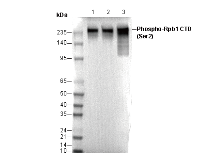 Lane 1: H-4-II-E
Lane 2: C2C12
Lane 3: COS-7