|
Cómo citar 1. Para citas en el texto (Materiales y métodos): 2. Para la tabla de recursos clave: |
||
|
Llamada gratuita: (877) 796-6397 -- Solo EE. UU. y Canadá -- |
Fax: +1-832-582-8590 Pedidos: +1-832-582-8158 |
Soporte técnico: +1-832-582-8158 Ext:3 Por favor, indique su número de pedido en el correo electrónico. Nos esforzamos por responder a todas las consultas por correo electrónico en el plazo de un día hábil. |
Descripción biológica
| Especificidad | Phospho-RIP3 (Ser232) Antibody [E19K17] reconoce los niveles endógenos de la proteína RIP3 total solo cuando está fosforilada en Ser232 |
|---|---|
| Antecedentes | La familia de proteínas quinasas de serina/treonina interactivas con receptores (RIP) —incluyendo RIP1, RIP2, RIP3 y RIP4— juega un papel crucial en la modulación de las respuestas celulares al estrés. Estas quinasas están involucradas en la iniciación de señales pro-supervivencia e inflamatorias, principalmente a través de la activación de NF-κB, así como en la promoción de vías de muerte celular como Apoptosis. Entre ellas, la receptor-interacting protein kinase 3 (RIPK3 o RIP3) es un actor central en la necroptosis, una forma regulada de muerte celular necrótica. RIP3 participa en cascadas de señalización iniciadas por el factor de necrosis tumoral (TNF), donde se asocia con RIP1 y el complejo del receptor de TNF para mediar tanto la activación de NF-κB como Apoptosis. Un evento clave en la necroptosis es la interacción entre RIP1 y RIP3, que forma la base del necrosoma, un complejo multiproteico que facilita la necrosis programada. Esta muerte celular de tipo necrótico se desencadena típicamente por la señalización de TNF en presencia de inhibición de caspasas. En humanos, la fosforilación de RIP3 en Serina 227 es crítica para su unión a la proteína tipo dominio de quinasa de linaje mixto (MLKL), lo que permite el ensamblaje del necrosoma. En ratones, la estimulación con TNF induce la fosforilación en Treonina 231 y Serina 232 en RIP3, lo cual es necesario para su interacción con MLKL de ratón. Cabe destacar que Ser-232 en RIP3 murina corresponde a Ser-227 en la proteína humana. Además, RIP1 y RIP3 contribuyen a la oligomerización del necrosoma, lo que promueve la formación de estructuras de señalización similares al amiloide esenciales para ejecutar la necroptosis. |
Información de uso
| Aplicación | WB, ELISA | Dilución |
|
||
|---|---|---|---|---|---|
| Reactividad | Mouse | ||||
| Fuente | Rabbit Monoclonal Antibody | MW | 53 kDa | ||
| Tampón de almacenamiento | PBS, pH 7.2+50% Glycerol+0.05% BSA+0.01% NaN3 | Almacenamiento (Desde la fecha de recepción) |
-20°C (avoid freeze-thaw cycles), 2 years | ||
| WB |
Experimental Protocol:
Sample preparation
1. Tissue: Lyse the tissue sample by adding an appropriate volume of ice-cold RIPA/Nuclear Lysis Buffer (containing Protease Inhibitor Cocktail, Phosphatase Inhibitor Cocktail),and homogenize the tissue at a low temperature. 2. Adherent cell: Aspirate the culture medium and wash the cells with ice-cold PBS twice. Lyse the cells by adding an appropriate volume of RIPA/Nuclear Lysis Buffer (containing Protease Inhibitor Cocktail, Phosphatase Inhibitor Cocktail) and put the sample on ice for 5 min. 3. Suspension cell: Transfer the culture medium to a pre-cooled centrifuge tube. Centrifuge and aspirate the supernatant. Wash the cells with ice-cold PBS twice. Lyse the cells by adding an appropriate volume of RIPA/Nuclear Lysis Buffer (containing Protease Inhibitor Cocktail, Phosphatase Inhibitor Cocktail) and put the sample on ice for 5 min. 4. Place the lysate into a pre-cooled microcentrifuge tube. Centrifuge at 4°C for 15 min. Collect the supernatant;
5. Remove a small volume of lysate to determine the protein concentration;
6. Combine the lysate with protein loading buffer. Boil 20 µL sample under 95-100°C for 5 min. Centrifuge for 5 min after cool down on ice.
Electrophoretic separation
1. According to the concentration of extracted protein, load appropriate amount of protein sample and marker onto SDS-PAGE gels for electrophoresis. Recommended separating gel (lower gel) concentration: 10%. Reference Table for Selecting SDS-PAGE Separation Gel Concentrations 2. Power up 80V for 30 minutes. Then the power supply is adjusted (110 V~150 V), the Marker is observed, and the electrophoresis can be stopped when the indicator band of the predyed protein Marker where the protein is located is properly separated. (Note that the current should not be too large when electrophoresis, too large current (more than 150 mA) will cause the temperature to rise, affecting the result of running glue. If high currents cannot be avoided, an ice bath can be used to cool the bath.)
Transfer membrane
1. Take out the converter, soak the clip and consumables in the pre-cooled converter;
2. Activate PVDF membrane with methanol for 1 min and rinse with transfer buffer;
3. Install it in the order of "black edge of clip - sponge - filter paper - filter paper - glue -PVDF membrane - filter paper - filter paper - sponge - white edge of clip"; 4. The protein was electrotransferred to PVDF membrane. ( 0.45 µm PVDF membrane is recommended ) Reference Table for Selecting PVDF Membrane Pore Size Specifications Recommended conditions for wet transfer: 200 mA, 120 min. ( Note that the transfer conditions can be adjusted according to the protein size. For high-molecular-weight proteins, a higher current and longer transfer time are recommended. However, ensure that the transfer tank remains at a low temperature to prevent gel melting.)
Block
1. After electrotransfer, wash the film with TBST at room temperature for 5 minutes;
2. Incubate the film in the blocking solution ( recommending 5% BSA solution)
for 1 hour at room temperature;
3. Wash the film with TBST for 3 times, 5 minutes each time.
Antibody incubation
1. Use 5% skim milk powder to prepare the primary antibody working liquid (recommended dilution ratio for primary antibody 1:1000), gently shake and incubate with the film at 4°C overnight; 2. Wash the film with TBST 3 times, 5 minutes each time;
3. Add the secondary antibody to the blocking solution and incubate with the film gently at room temperature for 1 hour;
4. After incubation, wash the film with TBST 3 times for 5 minutes each time.
Antibody staining
1. Add the prepared ECL luminescent substrate (or select other color developing substrate according to the second antibody) and mix evenly;
2. Incubate with the film for 1 minute, remove excess substrate (keep the film moist), wrap with plastic film, and expose in the imaging system.
|
Referencias
|
Datos de aplicación
WB
Validado por Selleck
-
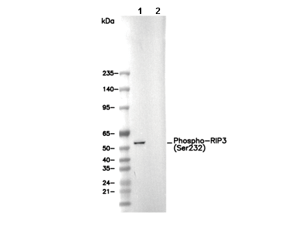 Lane 1: L-929, Lane 2: L-929 (phosphatase-treated)