|
Cómo citar 1. Para citas en el texto (Materiales y métodos): 2. Para la tabla de recursos clave: |
||
|
Llamada gratuita: (877) 796-6397 -- Solo EE. UU. y Canadá -- |
Fax: +1-832-582-8590 Pedidos: +1-832-582-8158 |
Soporte técnico: +1-832-582-8158 Ext:3 Por favor, indique su número de pedido en el correo electrónico. Nos esforzamos por responder a todas las consultas por correo electrónico en el plazo de un día hábil. |
Descripción biológica
| Especificidad | Phospho-Akt1 (Ser129) Antibody [K23C4] detecta niveles endógenos de la proteína Akt1 total solo cuando está fosforilada en Ser129. |
|---|---|
| Antecedentes | La Phospho-Akt1 (Ser129) es una modificación postraduccional específica de Akt1 (RAC-alfa serina/treonina-proteína quinasa, PKBα), que ocurre dentro de la región de unión flexible entre el dominio de homología de pleckstrina (PH) y el dominio quinasa de esta proteína de 480 aminoácidos. Akt1 comprende un dominio PH N-terminal para el reclutamiento de membrana mediado por PIP3, un dominio quinasa catalítico con sitios clave de fosforilación reguladora en Thr308 (bucle de activación) y Ser473 (motivo hidrofóbico), y una cola reguladora C-terminal. La fosforilación de Ser129 por la proteína quinasa CK2 genera un motivo consenso S-x-x-D/E que prepara la modificación posterior en Ser126 y estabiliza la conformación general de Akt1. Esta modificación hiperactiva Akt1 más allá de los efectos de la fosforilación canónica de PDK1/mTORC2 al fortalecer su asociación con el complejo chaperona HSP90, que protege a Thr308 de la desfosforilación mediada por PP2A, manteniendo así la actividad quinasa de Akt1. Además, la fosfo-Ser129 mejora la actividad transcripcional de β-catenina/TCF, ya sea directamente o estabilizando los componentes de la señalización Wnt, y bloquea a Akt1 en una conformación activa extendida que permite la selección de sustratos específicos de isoforma, como la fosforilación preferencial de la paladina, un proceso no reflejado en Akt2 debido a la ausencia del sitio equivalente Ser131. Esta modificación impulsa la remodelación citoesquelética, la supervivencia celular a través de la inhibición de Bad y FoxO, la proliferación al promover la estabilidad de la ciclina D1 y el secuestro de p27/p21, y la activación de mTORC1 a través de la fosforilación de TSC2, con una fosforilación jerárquica que asegura una amplificación robusta de la señal tras la estimulación por factores de crecimiento. En enfermedades, los niveles elevados de fosfo-Ser129 mejoran la supervivencia y la metástasis de las células cancerosas al amplificar la señalización oncogénica de PI3K/Akt (especialmente en cánceres de mama y próstata), contribuyen a la quimiorresistencia a través de una protección HSP90 sostenida y están implicados en trastornos metabólicos a través de la interrupción de la homeostasis de la glucosa al hiperactivar Akt1. |
Información de uso
| Aplicación | WB | Dilución |
|
||
|---|---|---|---|---|---|
| Reactividad | Human | ||||
| Fuente | Rabbit Monoclonal Antibody | MW | 55 kDa | ||
| Tampón de almacenamiento | PBS, pH 7.2+50% Glycerol+0.05% BSA+0.01% NaN3 | Almacenamiento (Desde la fecha de recepción) |
-20°C (avoid freeze-thaw cycles), 2 years | ||
| WB |
Experimental Protocol:
Sample preparation
1. Tissue: Lyse the tissue sample by adding an appropriate volume of ice-cold RIPA/NP-40 Lysis Buffer (containing Protease Inhibitor Cocktail, Phosphatase Inhibitor Cocktail),and homogenize the tissue at a low temperature. 2. Adherent cell: Aspirate the culture medium and wash the cells with ice-cold PBS twice. Lyse the cells by adding an appropriate volume of RIPA/NP-40 Lysis Buffer (containing Protease Inhibitor Cocktail, Phosphatase Inhibitor Cocktail) and put the sample on ice for 5 min. 3. Suspension cell: Transfer the culture medium to a pre-cooled centrifuge tube. Centrifuge and aspirate the supernatant. Wash the cells with ice-cold PBS twice. Lyse the cells by adding an appropriate volume of RIPA/NP-40 Lysis Buffer (containing Protease Inhibitor Cocktail, Phosphatase Inhibitor Cocktail) and put the sample on ice for 5 min. 4. Place the lysate into a pre-cooled microcentrifuge tube. Centrifuge at 4°C for 15 min. Collect the supernatant;
5. Remove a small volume of lysate to determine the protein concentration;
6. Combine the lysate with protein loading buffer. Boil 20 µL sample under 95-100°C for 5 min. Centrifuge for 5 min after cool down on ice.
Electrophoretic separation
1. According to the concentration of extracted protein, load appropriate amount of protein sample and marker onto SDS-PAGE gels for electrophoresis. Recommended separating gel (lower gel) concentration: 10%. Reference Table for Selecting SDS-PAGE Separation Gel Concentrations 2. Power up 80V for 30 minutes. Then the power supply is adjusted (110 V~150 V), the Marker is observed, and the electrophoresis can be stopped when the indicator band of the predyed protein Marker where the protein is located is properly separated. (Note that the current should not be too large when electrophoresis, too large current (more than 150 mA) will cause the temperature to rise, affecting the result of running glue. If high currents cannot be avoided, an ice bath can be used to cool the bath.)
Transfer membrane
1. Take out the converter, soak the clip and consumables in the pre-cooled converter;
2. Activate PVDF membrane with methanol for 1 min and rinse with transfer buffer;
3. Install it in the order of "black edge of clip - sponge - filter paper - filter paper - glue -PVDF membrane - filter paper - filter paper - sponge - white edge of clip"; 4. The protein was electrotransferred to PVDF membrane. ( 0.45 µm PVDF membrane is recommended ) Reference Table for Selecting PVDF Membrane Pore Size Specifications Recommended conditions for wet transfer: 200 mA, 120 min. ( Note that the transfer conditions can be adjusted according to the protein size. For high-molecular-weight proteins, a higher current and longer transfer time are recommended. However, ensure that the transfer tank remains at a low temperature to prevent gel melting.)
Block
1. After electrotransfer, wash the film with TBST at room temperature for 5 minutes;
2. Incubate the film in the blocking solution ( recommending 5% BSA solution)
for 1 hour at room temperature;
3. Wash the film with TBST for 3 times, 5 minutes each time.
Antibody incubation
1. Use 5% skim milk powder to prepare the primary antibody working liquid (recommended dilution ratio for primary antibody 1:1000), gently shake and incubate with the film at 4°C overnight; 2. Wash the film with TBST 3 times, 5 minutes each time;
3. Add the secondary antibody to the blocking solution and incubate with the film gently at room temperature for 1 hour;
4. After incubation, wash the film with TBST 3 times for 5 minutes each time.
Antibody staining
1. Add the prepared ECL luminescent substrate (or select other color developing substrate according to the second antibody) and mix evenly;
2. Incubate with the film for 1 minute, remove excess substrate (keep the film moist), wrap with plastic film, and expose in the imaging system.
|
Referencias
|
Datos de aplicación
WB
Validado por Selleck
-
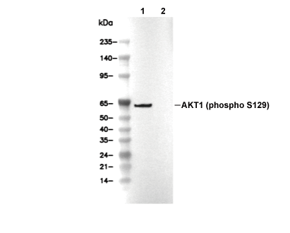 Lane 1: MCF-7, Lane 2: MCF-7 (Alkaline Phosphatase treated)