|
Cómo citar 1. Para citas en el texto (Materiales y métodos): 2. Para la tabla de recursos clave: |
||
|
Llamada gratuita: (877) 796-6397 -- Solo EE. UU. y Canadá -- |
Fax: +1-832-582-8590 Pedidos: +1-832-582-8158 |
Soporte técnico: +1-832-582-8158 Ext:3 Por favor, indique su número de pedido en el correo electrónico. Nos esforzamos por responder a todas las consultas por correo electrónico en el plazo de un día hábil. |
Descripción biológica
| Especificidad | MCM3 Antibody [B16A9] reconoce los niveles endógenos de la proteína MCM3 total. |
|---|---|
| Antecedentes | MCM3 es una subunidad central del complejo de mantenimiento de minicromosomas (MCM) 2–7, que actúa como la helicasa replicativa esencial para el inicio y la elongación de la replicación del DNA en células eucariotas. Esta gran proteína, de aproximadamente 971 aminoácidos, comparte una homología significativa con MCM2, especialmente en tres regiones conservadas críticas para la actividad de helicasa y la unión al DNA. Estructuralmente, MCM3 contiene múltiples sitios de fosforilación e interfaces que median las interacciones con otras subunidades MCM, particularmente MCM5, y se incorpora al anillo hexamérico MCM2-7 que se carga en los orígenes de replicación como parte del complejo pre-replicativo (pre-RC) durante las fases M tardía y G1. El reclutamiento de MCM3 a los orígenes depende del complejo de reconocimiento de orígenes (ORC), CDC6 y CDT1, asegurando que la licencia de replicación ocurra solo una vez por Cell Cycle. La actividad de MCM3 está estrictamente regulada por fosforilación, notablemente por quinasas dependientes de ciclina como la ciclina E/Cdk2, que fosforila la treonina 722 para promover la carga de la cromatina pero puede inhibir la replicación si está desregulada. También es un sustrato para las quinasas de punto de control ATM y ATR, vinculando MCM3 a las respuestas al daño del DNA y las vías de punto de control de la replicación. La acetilación por MCM3AP inhibe el inicio de la replicación del DNA y la progresión del Cell Cycle, añadiendo otra capa de control. La prolil isomerasa Pin1 modula la unión de MCM3 a la cromatina de una manera dependiente del Cell Cycle, integrando aún más la regulación postraduccional. La sobreexpresión de MCM3 de tipo salvaje puede causar un arresto en G1 y un retraso en la fase S, mientras que mutantes específicos como T722A pueden rescatar estos efectos. Aunque MCM3 es esencial para la progresión de la horquilla de replicación y la estabilidad del genoma, solo una fracción de los complejos cargados está activamente involucrada en la replicación, sirviendo los complejos en exceso como orígenes de respaldo y participando en la señalización del punto de control. MCM3 también interactúa con otras proteínas asociadas a la replicación y la cromatina, regulando la especificidad del origen y coordinando la replicación con la progresión del Cell Cycle. Las mutaciones o desequilibrios en la expresión de MCM3 pueden conducir a defectos en la licencia de replicación, inestabilidad genómica y arresto del Cell Cycle. |
Información de uso
| Aplicación | WB, IHC, FCM | Dilución |
|
||||||
|---|---|---|---|---|---|---|---|---|---|
| Reactividad | Human, Mouse, Rat | ||||||||
| Fuente | Rabbit Monoclonal Antibody | MW | 91 kDa | ||||||
| Tampón de almacenamiento | PBS, pH 7.2+50% Glycerol+0.05% BSA+0.01% NaN3 | Almacenamiento (Desde la fecha de recepción) |
-20°C (avoid freeze-thaw cycles), 2 years | ||||||
| WB |
Experimental Protocol:
Sample preparation
1. Tissue: Lyse the tissue sample by adding an appropriate volume of ice-cold RIPA/Nuclear Lysis Buffer (containing Protease Inhibitor Cocktail),and homogenize the tissue at a low temperature. 2. Adherent cell: Aspirate the culture medium and wash the cells with ice-cold PBS twice. Lyse the cells by adding an appropriate volume of RIPA/Nuclear Lysis Buffer (containing Protease Inhibitor Cocktail) and put the sample on ice for 5 min. 3. Suspension cell: Transfer the culture medium to a pre-cooled centrifuge tube. Centrifuge and aspirate the supernatant. Wash the cells with ice-cold PBS twice. Lyse the cells by adding an appropriate volume of RIPA/Nuclear Lysis Buffer (containing Protease Inhibitor Cocktail) and put the sample on ice for 5 min. 4. Place the lysate into a pre-cooled microcentrifuge tube. Centrifuge at 4°C for 15 min. Collect the supernatant;
5. Remove a small volume of lysate to determine the protein concentration;
6. Combine the lysate with protein loading buffer. Boil 20 µL sample under 95-100°C for 5 min. Centrifuge for 5 min after cool down on ice.
Electrophoretic separation
1. According to the concentration of extracted protein, load appropriate amount of protein sample and marker onto SDS-PAGE gels for electrophoresis. Recommended separating gel (lower gel) concentration: 10%. Reference Table for Selecting SDS-PAGE Separation Gel Concentrations 2. Power up 80V for 30 minutes. Then the power supply is adjusted (110 V~150 V), the Marker is observed, and the electrophoresis can be stopped when the indicator band of the predyed protein Marker where the protein is located is properly separated. (Note that the current should not be too large when electrophoresis, too large current (more than 150 mA) will cause the temperature to rise, affecting the result of running glue. If high currents cannot be avoided, an ice bath can be used to cool the bath.)
Transfer membrane
1. Take out the converter, soak the clip and consumables in the pre-cooled converter;
2. Activate PVDF membrane with methanol for 1 min and rinse with transfer buffer;
3. Install it in the order of "black edge of clip - sponge - filter paper - filter paper - glue -PVDF membrane - filter paper - filter paper - sponge - white edge of clip"; 4. The protein was electrotransferred to PVDF membrane. ( 0.45 µm PVDF membrane is recommended ) Reference Table for Selecting PVDF Membrane Pore Size Specifications Recommended conditions for wet transfer: 200 mA, 120 min. ( Note that the transfer conditions can be adjusted according to the protein size. For high-molecular-weight proteins, a higher current and longer transfer time are recommended. However, ensure that the transfer tank remains at a low temperature to prevent gel melting.)
Block
1. After electrotransfer, wash the film with TBST at room temperature for 5 minutes;
2. Incubate the film in the blocking solution for 1 hour at room temperature;
3. Wash the film with TBST for 3 times, 5 minutes each time.
Antibody incubation
1. Use 5% skim milk powder to prepare the primary antibody working liquid (recommended dilution ratio for primary antibody 1:1000), gently shake and incubate with the film at 4°C overnight; 2. Wash the film with TBST 3 times, 5 minutes each time;
3. Add the secondary antibody to the blocking solution and incubate with the film gently at room temperature for 1 hour;
4. After incubation, wash the film with TBST 3 times for 5 minutes each time.
Antibody staining
1. Add the prepared ECL luminescent substrate (or select other color developing substrate according to the second antibody) and mix evenly;
2. Incubate with the film for 1 minute, remove excess substrate (keep the film moist), wrap with plastic film, and expose in the imaging system.
|
| IHC |
Experimental Protocol:
Deparaffinization/Rehydration
1. Deparaffinize/hydrate sections:
2. Incubate sections in three washes of xylene for 5 min each.
3. Incubate sections in two washes of 100% ethanol for 10 min each.
4. Incubate sections in two washes of 95% ethanol for 10 min each.
5. Wash sections two times in dH2O for 5 min each.
6.Antigen retrieval: For Citrate: Heat slides in a microwave submersed in 1X citrate unmasking solution until boiling is initiated; continue with 10 min at a sub-boiling temperature (95°-98°C). Cool slides on bench top for 30 min.
Staining
1. Wash sections in dH2O three times for 5 min each.
2. Incubate sections in 3% hydrogen peroxide for 10 min.
3. Wash sections in dH2O two times for 5 min each.
4. Wash sections in wash buffer for 5 min.
5. Block each section with 100–400 µl of blocking solution for 1 hr at room temperature.
6. Remove blocking solution and add 100–400 µl primary antibody diluent in to each section. Incubate overnight at 4°C.
7. Remove antibody solution and wash sections with wash buffer three times for 5 min each.
8. Cover section with 1–3 drops HRPas needed. Incubate in a humidified chamber for 30 min at room temperature.
9. Wash sections three times with wash buffer for 5 min each.
10. Add DAB Chromogen Concentrate to DAB Diluent and mix well before use.
11. Apply 100–400 µl DAB to each section and monitor closely. 1–10 min generally provides an acceptable staining intensity.
12. Immerse slides in dH2O.
13. If desired, counterstain sections with hematoxylin.
14. Wash sections in dH2O two times for 5 min each.
15. Dehydrate sections: Incubate sections in 95% ethanol two times for 10 sec each; Repeat in 100% ethanol, incubating sections two times for 10 sec each; Repeat in xylene, incubating sections two times for 10 sec each.
16. Mount sections with coverslips and mounting medium.
|
Referencias
|
Datos de aplicación
WB
Validado por Selleck
-
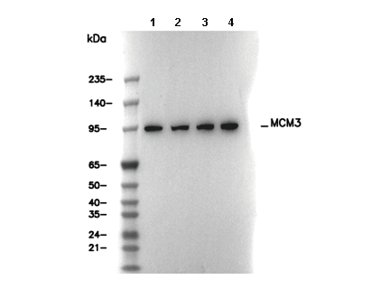 Lane 1: C6, Lane 2: RAW 264.7, Lane 3: PC-12, Lane 4: NIH/3T3