|
Cómo citar 1. Para citas en el texto (Materiales y métodos): 2. Para la tabla de recursos clave: |
||
|
Llamada gratuita: (877) 796-6397 -- Solo EE. UU. y Canadá -- |
Fax: +1-832-582-8590 Pedidos: +1-832-582-8158 |
Soporte técnico: +1-832-582-8158 Ext:3 Por favor, indique su número de pedido en el correo electrónico. Nos esforzamos por responder a todas las consultas por correo electrónico en el plazo de un día hábil. |
Descripción biológica
| Especificidad | Integrin β 3 Antibody [P12C15] reconoce los niveles endógenos de la proteína total Integrin β 3. |
|---|---|
| Antecedentes | Integrin β3 es una subunidad clave de la familia de receptores transmembrana Integrin que median la adhesión celular a la matriz extracelular (MEC) y transducen señales bidireccionalmente a través de la membrana plasmática. Comúnmente se empareja con las subunidades αV o αIIb para formar las integrinas αVβ3 y αIIbβ3, las cuales desempeñan roles esenciales en diversos procesos biológicos como la agregación plaquetaria (αIIbβ3), la angiogénesis, la migración celular y la supervivencia (αVβ3). Integrin β3 presenta un gran dominio extracelular, que incluye el dominio βA (similar a I) responsable de la unión al ligando, un dominio híbrido, un dominio de plexina-semaforina-Integrin (PSI) y múltiples dominios I-EGF, seguido de una única región transmembrana y una cola citoplasmática corta que interactúa con proteínas intracelulares. El dominio transmembrana es crítico para la heterodimerización α/β y la transmisión de señales. En su estado inactivo, la integrina β3 adopta una conformación doblada, pero al activarse, experimenta cambios conformacionales drásticos, extensión en la "rodilla" I-EGF1/2 y un balanceo del dominio híbrido, lo que permite la unión al ligando de alta afinidad y la separación de las "patas". La activación está estrechamente regulada por la señalización "de adentro hacia afuera", donde proteínas como la talina y la kindlina se unen a la cola citoplasmática, desencadenando cambios conformacionales de estados de baja a alta afinidad. La señalización "de afuera hacia adentro" sigue al compromiso del ligando, lo que lleva a la reorganización citoesquelética y la modulación de la expresión génica. La integrina β3 también contribuye a la resorción ósea por parte de los osteoclastos, el tráfico de células inmunes y la progresión tumoral. Su actividad es modulada por cationes divalentes (Ca²⁺, Mg²⁺, Mn²⁺), la fosforilación de residuos citoplasmáticos e interacciones con otras proteínas de membrana. Las variantes genéticas, como el polimorfismo Leu33Pro en el dominio PSI, están asociadas con respuestas inmunes alteradas y un mayor riesgo de trombosis. La desregulación de la integrina β3 está implicada en la trombosis, la metástasis del cáncer y las enfermedades inflamatorias. |
Información de uso
| Aplicación | WB, IP, FCM | Dilución |
|
||||||
|---|---|---|---|---|---|---|---|---|---|
| Reactividad | Human, Mouse, Rat | ||||||||
| Fuente | Rabbit Monoclonal Antibody | MW | 87 kDa | ||||||
| Tampón de almacenamiento | PBS, pH 7.2+50% Glycerol+0.05% BSA+0.01% NaN3 | Almacenamiento (Desde la fecha de recepción) |
-20°C (avoid freeze-thaw cycles), 2 years | ||||||
| WB |
Experimental Protocol:
Sample preparation
1. Tissue: Lyse the tissue sample by adding an appropriate volume of ice-cold RIPA/NP-40 Lysis Buffer (containing Protease Inhibitor Cocktail),and homogenize the tissue at a low temperature. 2. Adherent cell: Aspirate the culture medium and wash the cells with ice-cold PBS twice. Lyse the cells by adding an appropriate volume of RIPA/NP-40 Lysis Buffer (containing Protease Inhibitor Cocktail) and put the sample on ice for 5 min. 3. Suspension cell: Transfer the culture medium to a pre-cooled centrifuge tube. Centrifuge and aspirate the supernatant. Wash the cells with ice-cold PBS twice. Lyse the cells by adding an appropriate volume of RIPA/NP-40 Lysis Buffer (containing Protease Inhibitor Cocktail) and put the sample on ice for 5 min. 4. Place the lysate into a pre-cooled microcentrifuge tube. Centrifuge at 4°C for 15 min. Collect the supernatant;
5. Remove a small volume of lysate to determine the protein concentration;
6. Combine the lysate with protein loading buffer. Boil 20 µL sample under 95-100°C for 5 min. Centrifuge for 5 min after cool down on ice.
Electrophoretic separation
1. According to the concentration of extracted protein, load appropriate amount of protein sample and marker onto SDS-PAGE gels for electrophoresis. Recommended separating gel (lower gel) concentration: 10%. Reference Table for Selecting SDS-PAGE Separation Gel Concentrations 2. Power up 80V for 30 minutes. Then the power supply is adjusted (110 V~150 V), the Marker is observed, and the electrophoresis can be stopped when the indicator band of the predyed protein Marker where the protein is located is properly separated. (Note that the current should not be too large when electrophoresis, too large current (more than 150 mA) will cause the temperature to rise, affecting the result of running glue. If high currents cannot be avoided, an ice bath can be used to cool the bath.)
Transfer membrane
1. Take out the converter, soak the clip and consumables in the pre-cooled converter;
2. Activate PVDF membrane with methanol for 1 min and rinse with transfer buffer;
3. Install it in the order of "black edge of clip - sponge - filter paper - filter paper - glue -PVDF membrane - filter paper - filter paper - sponge - white edge of clip"; 4. The protein was electrotransferred to PVDF membrane. ( 0.45 µm PVDF membrane is recommended ) Reference Table for Selecting PVDF Membrane Pore Size Specifications Recommended conditions for wet transfer: 200 mA, 120 min. ( Note that the transfer conditions can be adjusted according to the protein size. For high-molecular-weight proteins, a higher current and longer transfer time are recommended. However, ensure that the transfer tank remains at a low temperature to prevent gel melting.)
Block
1. After electrotransfer, wash the film with TBST at room temperature for 5 minutes;
2. Incubate the film in the blocking solution for 1 hour at room temperature;
3. Wash the film with TBST for 3 times, 5 minutes each time.
Antibody incubation
1. Use 5% skim milk powder to prepare the primary antibody working liquid (recommended dilution ratio for primary antibody 1:1000), gently shake and incubate with the film at 4°C overnight; 2. Wash the film with TBST 3 times, 5 minutes each time;
3. Add the secondary antibody to the blocking solution and incubate with the film gently at room temperature for 1 hour;
4. After incubation, wash the film with TBST 3 times for 5 minutes each time.
Antibody staining
1. Add the prepared ECL luminescent substrate (or select other color developing substrate according to the second antibody) and mix evenly;
2. Incubate with the film for 1 minute, remove excess substrate (keep the film moist), wrap with plastic film, and expose in the imaging system.
|
Referencias
|
Datos de aplicación
WB
Validado por Selleck
-
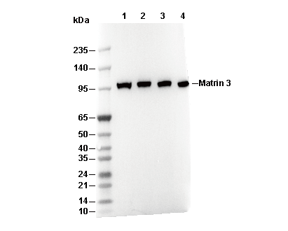 Lane 1: U-87MG, Lane 2: HEL, Lane 3: HUVEC, Lane 4: C2C12