|
Cómo citar 1. Para citas en el texto (Materiales y métodos): 2. Para la tabla de recursos clave: |
||
|
Llamada gratuita: (877) 796-6397 -- Solo EE. UU. y Canadá -- |
Fax: +1-832-582-8590 Pedidos: +1-832-582-8158 |
Soporte técnico: +1-832-582-8158 Ext:3 Por favor, indique su número de pedido en el correo electrónico. Nos esforzamos por responder a todas las consultas por correo electrónico en el plazo de un día hábil. |
Descripción biológica
| Especificidad | HACE1 Antibody [A17B18] reconoce los niveles endógenos de la proteína HACE1 total. |
|---|---|
| Antecedentes | El gen HACE1 (HECT domain and ankyrin repeat-containing E3 ubiquitin-protein ligase 1) está localizado en el cromosoma 6 y codifica una proteína compuesta por 909 aminoácidos. HACE1 se expresa fuertemente en varios tejidos humanos, incluyendo el corazón, cerebro, placenta, páncreas, y riñones tanto fetales como adultos. El ARNm de HACE1 se expresa ubicuamente en tejidos humanos normales. Los niveles reducidos de expresión de HACE1 están fuertemente asociados con la hipermetilación de dos islas CpG localizadas corriente arriba de su locus génico, sugiriendo que la regulación epigenética juega un papel en su silenciamiento. HACE1 está frecuentemente regulado a la baja en varios tipos de cáncer, incluyendo el linfoma de células T/natural killer (NKTCL), cáncer colorrectal y cáncer gástrico. Subcelularmente, HACE1 se localiza principalmente en el retículo endoplasmático y el citosol, aunque típicamente solo se detectan bajos niveles de proteína endógena. Funcionalmente, HACE1 actúa como una E3 Ligase y se asocia con la enzima E2 UBCH7 para catalizar la ubiquitinación de proteínas objetivo. Está notablemente implicado en la degradación dependiente de fosforilación de la ciclina D1, jugando así un papel en la inhibición de la progresión del ciclo celular. HACE1 también se une preferentemente a la forma activa, unida a GTP, de la pequeña GTPasa Rac1 y promueve su poliubiquitinación. Esta actividad es esencial para la degradación de Rac1 en respuesta al factor necrotizante citotóxico 1 (CNF1), facilitando la invasión bacteriana de monocapas de células endoteliales, y resaltando un papel para HACE1 en los mecanismos de defensa inmune innata. La pérdida de HACE1 conduce al desarrollo espontáneo de tumores de aparición tardía, apoyando aún más su función como supresor tumoral. El gen se encuentra dentro de la región cromosómica 6q21, que es un punto caliente implicado en múltiples cánceres humanos, subrayando su importancia clínica en la tumorigénesis. |
Información de uso
| Aplicación | WB | Dilución |
|
||
|---|---|---|---|---|---|
| Reactividad | Mouse, Rat, Human | ||||
| Fuente | Rabbit Monoclonal Antibody | MW | 102 kDa | ||
| Tampón de almacenamiento | PBS, pH 7.2+50% Glycerol+0.05% BSA+0.01% NaN3 | Almacenamiento (Desde la fecha de recepción) |
-20°C (avoid freeze-thaw cycles), 2 years | ||
| WB |
Experimental Protocol:
Sample preparation
1. Tissue: Lyse the tissue sample by adding an appropriate volume of ice-cold RIPA/NP-40 Lysis Buffer (containing Protease Inhibitor Cocktail),and homogenize the tissue at a low temperature. 2. Adherent cell: Aspirate the culture medium and wash the cells with ice-cold PBS twice. Lyse the cells by adding an appropriate volume of RIPA/NP-40 Lysis Buffer (containing Protease Inhibitor Cocktail) and put the sample on ice for 5 min. 3. Suspension cell: Transfer the culture medium to a pre-cooled centrifuge tube. Centrifuge and aspirate the supernatant. Wash the cells with ice-cold PBS twice. Lyse the cells by adding an appropriate volume of RIPA/NP-40 Lysis Buffer (containing Protease Inhibitor Cocktail) and put the sample on ice for 5 min. 4. Place the lysate into a pre-cooled microcentrifuge tube. Centrifuge at 4°C for 15 min. Collect the supernatant;
5. Remove a small volume of lysate to determine the protein concentration;
6. Combine the lysate with protein loading buffer. Boil 20 µL sample under 95-100°C for 5 min. Centrifuge for 5 min after cool down on ice.
Electrophoretic separation
1. According to the concentration of extracted protein, load appropriate amount of protein sample and marker onto SDS-PAGE gels for electrophoresis. Recommended separating gel (lower gel) concentration: 5%. Reference Table for Selecting SDS-PAGE Separation Gel Concentrations 2. Power up 80V for 30 minutes. Then the power supply is adjusted (110 V~150 V), the Marker is observed, and the electrophoresis can be stopped when the indicator band of the predyed protein Marker where the protein is located is properly separated. (Note that the current should not be too large when electrophoresis, too large current (more than 150 mA) will cause the temperature to rise, affecting the result of running glue. If high currents cannot be avoided, an ice bath can be used to cool the bath.)
Transfer membrane
1. Take out the converter, soak the clip and consumables in the pre-cooled converter;
2. Activate PVDF membrane with methanol for 1 min and rinse with transfer buffer;
3. Install it in the order of "black edge of clip - sponge - filter paper - filter paper - glue -PVDF membrane - filter paper - filter paper - sponge - white edge of clip"; 4. The protein was electrotransferred to PVDF membrane. ( 0.45 µm PVDF membrane is recommended ) Reference Table for Selecting PVDF Membrane Pore Size Specifications Recommended conditions for wet transfer: 200 mA, 120 min. ( Note that the transfer conditions can be adjusted according to the protein size. For high-molecular-weight proteins, a higher current and longer transfer time are recommended. However, ensure that the transfer tank remains at a low temperature to prevent gel melting.)
Block
1. After electrotransfer, wash the film with TBST at room temperature for 5 minutes;
2. Incubate the film in the blocking solution for 1 hour at room temperature;
3. Wash the film with TBST for 3 times, 5 minutes each time.
Antibody incubation
1. Use 5% skim milk powder to prepare the primary antibody working liquid (recommended dilution ratio for primary antibody 1:1000), gently shake and incubate with the film at 4°C overnight; 2. Wash the film with TBST 3 times, 5 minutes each time;
3. Add the secondary antibody to the blocking solution and incubate with the film gently at room temperature for 1 hour;
4. After incubation, wash the film with TBST 3 times for 5 minutes each time.
Antibody staining
1. Add the prepared ECL luminescent substrate (or select other color developing substrate according to the second antibody) and mix evenly;
2. Incubate with the film for 1 minute, remove excess substrate (keep the film moist), wrap with plastic film, and expose in the imaging system.
|
Referencias
|
Datos de aplicación
WB
Validado por Selleck
-
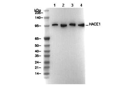 Lane 1: HEK293, Lane 2: SH-SY5Y, Lane 3: Mouse brain, Lane 4: Rat brain