|
Cómo citar 1. Para citas en el texto (Materiales y métodos): 2. Para la tabla de recursos clave: |
||
|
Llamada gratuita: (877) 796-6397 -- Solo EE. UU. y Canadá -- |
Fax: +1-832-582-8590 Pedidos: +1-832-582-8158 |
Soporte técnico: +1-832-582-8158 Ext:3 Por favor, indique su número de pedido en el correo electrónico. Nos esforzamos por responder a todas las consultas por correo electrónico en el plazo de un día hábil. |
Descripción biológica
| Especificidad | Glutathione Antibody [A11G7] detecta niveles endógenos de proteína total de Glutathione. |
|---|---|
| Antecedentes | Glutathione (GSH) es un tripéptido tiol antioxidante de bajo peso molecular compuesto por glutamato, cisteína y glicina, sintetizado a través de dos pasos secuenciales dependientes de ATP catalizados por la glutamato-cisteína ligasa y la glutatión sintetasa. Existe principalmente en formas reducida (GSH) y disulfuro oxidada (GSSG), que se interconvierten para regular el equilibrio redox celular. El GSH presenta un enlace peptídico γ-glutamil único entre el γ-carboxilo del glutamato y el grupo amino de la cisteína, lo que le confiere resistencia a la degradación por la γ-glutamil ciclotransferasa y la mayoría de las peptidasas; el tiol de la cisteína (-SH) actúa como centro nucleofílico para la química redox, mientras que la glicina estabiliza el C-terminal. El GSH elimina directamente las especies reactivas de oxígeno y nitrógeno (ROS/RNS) a través del intercambio tiol-disulfuro, sirve como un reductor esencial para las glutatión peroxidasas (GPx) que desintoxican los peróxidos (con el GSSG reducido de nuevo a GSH por la glutatión reductasa utilizando NADPH), y conjuga xenobióticos electrófilos, metales pesados y toxinas endógenas a través de las glutatión S-transferasas (GSTs) para la desintoxicación de fase II y la exportación de ácido mercaptúrico. Estos procesos mantienen la homeostasis de los sulfhidrilos proteicos, apoyan la expresión génica antioxidante impulsada por Nrf2, facilitan el ensamblaje de clústeres de hierro-azufre y el tráfico de metales, y modulan vías de señalización como NF-κB. El agotamiento de GSH está implicado en enfermedades relacionadas con el estrés oxidativo, incluyendo la enfermedad de Parkinson, la cirrosis hepática, el cáncer, la diabetes y el envejecimiento prematuro. |
Información de uso
| Aplicación | IF, FCM | Dilución |
|
||||
|---|---|---|---|---|---|---|---|
| Reactividad | |||||||
| Fuente | Mouse Monoclonal Antibody | MW | |||||
| Tampón de almacenamiento | PBS, pH 7.2+50% Glycerol+0.05% BSA+0.01% NaN3 | Almacenamiento (Desde la fecha de recepción) |
-20°C (avoid freeze-thaw cycles), 2 years | ||||
| IF |
Experimental Protocol:
Sample Preparation
1. Adherent Cells: Place a clean, sterile coverslip in a culture dish. Once the cells grow to near confluence as a monolayer, remove the coverslip for further use.
2. Suspension Cells: Seed the cells onto a clean, sterile slide coated with poly-L-lysine.
3. Frozen Sections: Allow the slide to thaw at room temperature. Wash it with pure water or PBS for 2 times, 3 minutes each time.
4. Paraffin Sections: Deparaffinization and rehydration. Wash the slide with pure water or PBS for 3 times, 3 minutes each time. Then perform antigen retrieval.
Fixation
1. Fix the cell coverslips/spots or tissue sections at room temperature using a fixative such as 4% paraformaldehyde (4% PFA) for 10-15 minutes.
2. Wash the sample with PBS for 3 times, 3 minutes each time.
Permeabilization
1.Add a detergent such as 0.1–0.3% Triton X-100 to the sample and incubate at room temperature for 10–20 minutes.
(Note: This step is only required for intracellular antigens. For antigens expressed on the cell membrane, this step is unnecessary.)
Wash the sample with PBS for 3 times, 3 minutes each time.
Blocking
Add blocking solution and incubate at room temperature for at least 1 hour. (Common blocking solutions include: serum from the same source as the secondary antibody, BSA, or goat serum.)
Note: Ensure the sample remains moist during and after the blocking step to prevent drying, which can lead to high background.
Immunofluorescence Staining (Day 1)
1. Remove the blocking solution and add the diluted primary antibody.
2. Incubate the sample in a humidified chamber at 4°C overnight.
Immunofluorescence Staining (Day 2)
1. Remove the primary antibody and wash with PBST for 3 times, 5 minutes each time.
2. Add the diluted fluorescent secondary antibody and incubate in the dark at 4°C for 1–2 hours.
3. Remove the secondary antibody and wash with PBST for 3 times, 5 minutes each time.
4. Add diluted DAPI and incubate at room temperature in the dark for 5–10 minutes.
5. Wash with PBST for 3 times, 5 minutes each time.
Mounting
1. Mount the sample with an anti-fade mounting medium.
2. Allow the slide to dry at room temperature overnight in the dark.
3. Store the slide in a slide storage box at 4°C, protected from light.
|
Referencias
|
Datos de aplicación
IF
Validado por Selleck
-
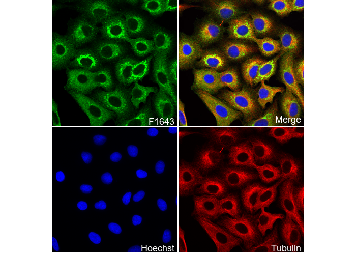 Immunofluorescent analysis of A549 cells using F1643 (green, 1:100), Hoechst (blue) and tubulin (Red).