|
Cómo citar 1. Para citas en el texto (Materiales y métodos): 2. Para la tabla de recursos clave: |
||
|
Llamada gratuita: (877) 796-6397 -- Solo EE. UU. y Canadá -- |
Fax: +1-832-582-8590 Pedidos: +1-832-582-8158 |
Soporte técnico: +1-832-582-8158 Ext:3 Por favor, indique su número de pedido en el correo electrónico. Nos esforzamos por responder a todas las consultas por correo electrónico en el plazo de un día hábil. |
Descripción biológica
| Especificidad | EWSR1/EWS Antibody [K7J15] reconoce los niveles endógenos de la proteína EWSR1/EWS total. |
|---|---|
| Antecedentes | EWSR1 (Ewing Sarcoma RNA-binding protein 1) es una proteína multifuncional codificada por el gen EWSR1 ubicado en el cromosoma 22q12.2, que desempeña papeles críticos en la transcripción, el empalme de ARN y la estabilidad genómica. Contiene un dominio de transactivación N-terminal (TAD) que interactúa con factores de transcripción como TFIID y la ARN polimerasa II, un dominio de unión a ARN (RBD) para la unión de ARN y múltiples dominios de baja complejidad (LCD) que facilitan interacciones dinámicas con ácidos nucleicos y proteínas. EWSR1 existe en dos modalidades visuales dentro del nucleoplasma: un estado distribuido y focos concentrados, que colocalizan con la ARN polimerasa II fosforilada en regiones eucromáticas para regular la transcripción activa. EWSR1 resuelve los bucles R —estructuras de ácidos nucleicos de tres hebras— a través de su RBD y su dominio de dedo de zinc para prevenir el estrés transcripcional y la inestabilidad genómica. En el sarcoma de Ewing, EWSR1 forma proteínas de fusión como EWS-FLI1, que alteran los estados de la cromatina al reclutar histonas metiltransferasas y acetiltransferasas para activar oncogenes como c-Myc mientras reprimen los supresores tumorales como p53. Estas proteínas de fusión también interactúan con regiones potenciadoras y repeticiones microsatélite para impulsar programas transcripcionales oncogénicos. La pérdida de función o las mutaciones de EWSR1 conducen a una meiosis deteriorada, envejecimiento prematuro, densidad ósea reducida y disfunción inmunitaria. |
Información de uso
| Aplicación | WB, IHC, IF, FCM | Dilución |
|
||||||||
|---|---|---|---|---|---|---|---|---|---|---|---|
| Reactividad | Human, Mouse, Rat | ||||||||||
| Fuente | Rabbit Monoclonal Antibody | MW | 68 kDa | ||||||||
| Tampón de almacenamiento | PBS, pH 7.2+50% Glycerol+0.05% BSA+0.01% NaN3 | Almacenamiento (Desde la fecha de recepción) |
-20°C (avoid freeze-thaw cycles), 2 years | ||||||||
| WB |
Experimental Protocol:
Sample preparation
1. Tissue: Lyse the tissue sample by adding an appropriate volume of ice-cold RIPA/NP-40 Lysis Buffer (containing Protease Inhibitor Cocktail),and homogenize the tissue at a low temperature or lyse it by sonication on ice, then incubate on ice for 30 minutes. 2. Adherent cell: Aspirate the culture medium and transfer the cells into an EP tube. Wash the cells with ice-cold PBS twice. Add an appropriate volume of RIPA/NP-40 Lysis Buffer (containing Protease Inhibitor Cocktail), sonicate to lyse the cells, and incubate on ice for 30 minutes. 3. Suspension cell: Transfer the culture medium to a pre-cooled centrifuge tube. Centrifuge and aspirate the supernatant. Wash the cells with ice-cold PBS twice.Add an appropriate volume of RIPA/NP-40 Lysis Buffer (containing Protease Inhibitor Cocktail), sonicate to lyse the cells, and incubate on ice for 30 minutes. 4. Place the lysate into a pre-cooled microcentrifuge tube. Centrifuge at 4°C for 15 min. Collect the supernatant;
5. Remove a small volume of lysate to determine the protein concentration;
6. Combine the lysate with protein loading buffer. Boil 20 µL sample under 95-100°C for 5 min. Centrifuge for 5 min after cool down on ice.
Electrophoretic separation
1. According to the concentration of extracted protein, load appropriate amount of protein sample and marker onto SDS-PAGE gels for electrophoresis. Recommended separating gel (lower gel) concentration: 10%. Reference Table for Selecting SDS-PAGE Separation Gel Concentrations 2. Power up 80V for 30 minutes. Then the power supply is adjusted (110 V~150 V), the Marker is observed, and the electrophoresis can be stopped when the indicator band of the predyed protein Marker where the protein is located is properly separated. (Note that the current should not be too large when electrophoresis, too large current (more than 150 mA) will cause the temperature to rise, affecting the result of running glue. If high currents cannot be avoided, an ice bath can be used to cool the bath.)
Transfer membrane
1. Take out the converter, soak the clip and consumables in the pre-cooled converter;
2. Activate PVDF membrane with methanol for 1 min and rinse with transfer buffer;
3. Install it in the order of "black edge of clip - sponge - filter paper - filter paper - glue -PVDF membrane - filter paper - filter paper - sponge - white edge of clip"; 4. The protein was electrotransferred to PVDF membrane. ( 0.45 µm PVDF membrane is recommended ) Reference Table for Selecting PVDF Membrane Pore Size Specifications Recommended conditions for wet transfer: 200 mA, 120 min. ( Note that the transfer conditions can be adjusted according to the protein size. For high-molecular-weight proteins, a higher current and longer transfer time are recommended. However, ensure that the transfer tank remains at a low temperature to prevent gel melting.)
Block
1. After electrotransfer, wash the film with TBST at room temperature for 5 minutes;
2. Incubate the film in the blocking solution for 1 hour at room temperature;
3. Wash the film with TBST for 3 times, 5 minutes each time.
Antibody incubation
1. Use 5% skim milk powder to prepare the primary antibody working liquid (recommended dilution ratio for primary antibody 1:100000), gently shake and incubate with the film at 4°C overnight; 2. Wash the film with TBST 3 times, 5 minutes each time;
3. Add the secondary antibody to the blocking solution and incubate with the film gently at room temperature for 1 hour;
4. After incubation, wash the film with TBST 3 times for 5 minutes each time.
Antibody staining
1389. Add the prepared ECL luminescent substrate (or select other color developing substrate according to the second antibody) and mix evenly;
2. Incubate with the film for 1 minute, remove excess substrate (keep the film moist), wrap with plastic film, and expose in the imaging system.
|
Referencias
|
Datos de aplicación
WB
Validado por Selleck
-
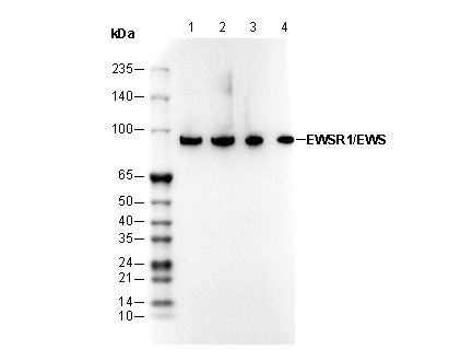 Lane 1: K562
Lane 2: HeLa
Lane 3: Jurkat
Lane 4: F9
IF
Validado por Selleck
-
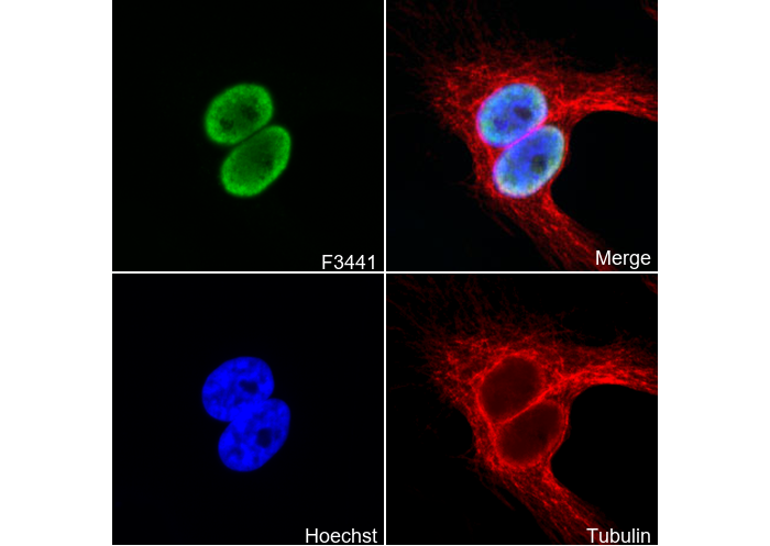 Immunofluorescent analysis of Hela cells using F3441 (green, 1:500), Hoechst (blue) and tubulin (Red).