|
Cómo citar 1. Para citas en el texto (Materiales y métodos): 2. Para la tabla de recursos clave: |
||
|
Llamada gratuita: (877) 796-6397 -- Solo EE. UU. y Canadá -- |
Fax: +1-832-582-8590 Pedidos: +1-832-582-8158 |
Soporte técnico: +1-832-582-8158 Ext:3 Por favor, indique su número de pedido en el correo electrónico. Nos esforzamos por responder a todas las consultas por correo electrónico en el plazo de un día hábil. |
Descripción biológica
| Especificidad | EF1α Antibody [M10N21] detecta los niveles endógenos de proteína EF1α total. |
|---|---|
| Antecedentes | El factor de elongación 1 alfa (EF1α) existe como dos isoformas estrechamente relacionadas, EF1α1 y EF1α2, que comparten más del 90% de identidad de secuencia y desempeñan roles equivalentes en la síntesis de proteínas al mediar la entrega dependiente de GTP de aminoacil-tRNAs al ribosoma. A pesar de su similitud funcional, las dos isoformas exhiben perfiles de expresión distintos. EF1α1 se expresa ampliamente en tejidos como el cerebro, la placenta, el pulmón, el hígado, el riñón y el páncreas, mientras que la expresión de EF1α2 está en gran medida restringida al cerebro, el corazón y el músculo esquelético. La desregulación específica de isoformas ha sido implicada en la biología del cáncer. En el tejido mamario normal, EF1α1 se expresa en niveles relativamente altos, mientras que EF1α2 es mínimamente detectable. Sin embargo, en el carcinoma de mama, ambas isoformas están reguladas al alza, y la sobreexpresión de EF1α2 parece tener mayor importancia biológica para el desarrollo y la progresión tumoral. Más allá de su papel canónico en la traducción, EF1α también funciona como una proteína que interactúa con pAkt, regulando la estabilidad y la señalización de Akt fosforilada, lo que lo vincula a las vías oncogénicas. Clínicamente, las isoformas de EF1α se han asociado con respuestas autoinmunes. EF1α1, en particular, ha sido identificado como un autoantígeno en aproximadamente el 66% de los pacientes con síndrome de Felty, destacando su potencial relevancia inmunológica. Colectivamente, la familia EF1α representa un grupo multifuncional de proteínas con roles clave en la traducción, la oncogénesis y la regulación inmunitaria. |
Información de uso
| Aplicación | WB, IP | Dilución |
|
||||
|---|---|---|---|---|---|---|---|
| Reactividad | Mammals | ||||||
| Fuente | Mouse Monoclonal Antibody | MW | 53 kDa | ||||
| Tampón de almacenamiento | PBS, pH 7.2+50% Glycerol+0.05% BSA+0.01% NaN3 | Almacenamiento (Desde la fecha de recepción) |
-20°C (avoid freeze-thaw cycles), 2 years | ||||
| WB |
Experimental Protocol:
Sample preparation
1. Tissue: Lyse the tissue sample by adding an appropriate volume of ice-cold RIPA/NP-40 Lysis Buffer (containing Protease Inhibitor Cocktail),and homogenize the tissue at a low temperature. 2. Adherent cell: Aspirate the culture medium and wash the cells with ice-cold PBS twice. Lyse the cells by adding an appropriate volume of RIPA/NP-40 Lysis Buffer (containing Protease Inhibitor Cocktail) and put the sample on ice for 5 min. 3. Suspension cell: Transfer the culture medium to a pre-cooled centrifuge tube. Centrifuge and aspirate the supernatant. Wash the cells with ice-cold PBS twice. Lyse the cells by adding an appropriate volume of RIPA/NP-40 Lysis Buffer (containing Protease Inhibitor Cocktail) and put the sample on ice for 5 min. 4. Place the lysate into a pre-cooled microcentrifuge tube. Centrifuge at 4°C for 15 min. Collect the supernatant;
5. Remove a small volume of lysate to determine the protein concentration;
6. Combine the lysate with protein loading buffer. Boil 20 µL sample under 95-100°C for 5 min. Centrifuge for 5 min after cool down on ice.
Electrophoretic separation
1. According to the concentration of extracted protein, load appropriate amount of protein sample and marker onto SDS-PAGE gels for electrophoresis. Recommended separating gel (lower gel) concentration: 10%. Reference Table for Selecting SDS-PAGE Separation Gel Concentrations 2. Power up 80V for 30 minutes. Then the power supply is adjusted (110 V~150 V), the Marker is observed, and the electrophoresis can be stopped when the indicator band of the predyed protein Marker where the protein is located is properly separated. (Note that the current should not be too large when electrophoresis, too large current (more than 150 mA) will cause the temperature to rise, affecting the result of running glue. If high currents cannot be avoided, an ice bath can be used to cool the bath.)
Transfer membrane
1. Take out the converter, soak the clip and consumables in the pre-cooled converter;
2. Activate PVDF membrane with methanol for 1 min and rinse with transfer buffer;
3. Install it in the order of "black edge of clip - sponge - filter paper - filter paper - glue -PVDF membrane - filter paper - filter paper - sponge - white edge of clip"; 4. The protein was electrotransferred to PVDF membrane. ( 0.45 µm PVDF membrane is recommended ) Reference Table for Selecting PVDF Membrane Pore Size Specifications Recommended conditions for wet transfer: 200 mA, 120 min. ( Note that the transfer conditions can be adjusted according to the protein size. For high-molecular-weight proteins, a higher current and longer transfer time are recommended. However, ensure that the transfer tank remains at a low temperature to prevent gel melting.)
Block
1. After electrotransfer, wash the film with TBST at room temperature for 5 minutes;
2. Incubate the film in the blocking solution for 1 hour at room temperature;
3. Wash the film with TBST for 3 times, 5 minutes each time.
Antibody incubation
1. Use 5% skim milk powder to prepare the primary antibody working liquid (recommended dilution ratio for primary antibody 1:250), gently shake and incubate with the film at 4°C overnight; 2. Wash the film with TBST 3 times, 5 minutes each time;
3. Add the secondary antibody to the blocking solution and incubate with the film gently at room temperature for 1 hour;
4. After incubation, wash the film with TBST 3 times for 5 minutes each time.
Antibody staining
1. Add the prepared ECL luminescent substrate (or select other color developing substrate according to the second antibody) and mix evenly;
2. Incubate with the film for 1 minute, remove excess substrate (keep the film moist), wrap with plastic film, and expose in the imaging system.
|
Referencias
|
Datos de aplicación
WB
Validado por Selleck
-
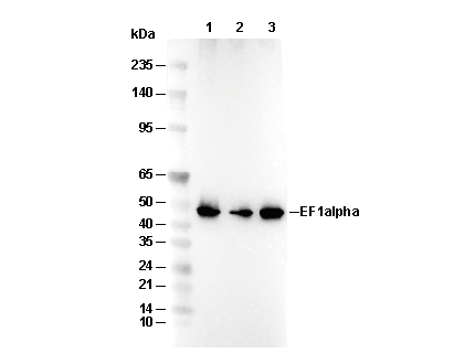 Lane 1: Jurkat, Lane 2: A431, Lane 3: 3T3