|
Cómo citar 1. Para citas en el texto (Materiales y métodos): 2. Para la tabla de recursos clave: |
||
|
Llamada gratuita: (877) 796-6397 -- Solo EE. UU. y Canadá -- |
Fax: +1-832-582-8590 Pedidos: +1-832-582-8158 |
Soporte técnico: +1-832-582-8158 Ext:3 Por favor, indique su número de pedido en el correo electrónico. Nos esforzamos por responder a todas las consultas por correo electrónico en el plazo de un día hábil. |
Descripción biológica
| Especificidad | DnaK Antibody [E21A24] detecta los niveles endógenos de la proteína DnaK total. |
|---|---|
| Antecedentes | DnaK es una proteína de choque térmico bacteriana altamente conservada que pertenece a la familia HSP70, estudiada principalmente en Escherichia coli, donde funciona como una chaperona molecular crucial para el plegamiento de proteínas, el replegamiento de proteínas desnaturalizadas y la homeostasis de proteínas bajo condiciones de estrés. DnaK consta de dos dominios principales: un dominio de unión a nucleótidos (NBD) N-terminal de 44 kDa responsable de la unión y la hidrólisis de ATP, y un dominio de unión a sustrato (SBD) C-terminal de 25 kDa, que incluye un subdominio en sándwich β que une sustratos proteicos desplegados a través de parches hidrofóbicos expuestos. DnaK opera a través de un ciclo dependiente de ATP donde la unión de ATP induce una conformación abierta que permite la liberación del sustrato, mientras que la hidrólisis de ATP desencadena una conformación cerrada que conduce a una unión de alta afinidad al sustrato. DnaK forma dímeros transitorios en su estado unido a ATP, lo que mejora su interacción con la cochaperona DnaJ (Hsp40), facilitando el reconocimiento y la entrega eficiente del sustrato. La cochaperona GrpE actúa como un factor de intercambio de nucleótidos, acelerando la liberación de ADP y reiniciando DnaK para otro ciclo. DnaK ayuda a prevenir la agregación de proteínas durante el estrés celular y participa en procesos críticos como la replicación del ADN, mediada por interacciones con la cochaperona DnaJ y otros socios. Esta red de chaperonas es esencial para la supervivencia bajo choque térmico y otros estreses proteotóxicos, manteniendo el control de calidad de las proteínas y la homeostasis celular. |
Información de uso
| Aplicación | WB | Dilución |
|
||
|---|---|---|---|---|---|
| Reactividad | Escherichia coli | ||||
| Fuente | Mouse Monoclonal Antibody | MW | 70 kDa | ||
| Tampón de almacenamiento | PBS, pH 7.2+50% Glycerol+0.05% BSA+0.01% NaN3 | Almacenamiento (Desde la fecha de recepción) |
-20°C (avoid freeze-thaw cycles), 2 years | ||
| WB |
Experimental Protocol:
Sample preparation
1. Tissue: Lyse the tissue sample by adding an appropriate volume of ice-cold RIPA/NP-40 Lysis Buffer (containing Protease Inhibitor Cocktail),and homogenize the tissue at a low temperature. 2. Adherent cell: Aspirate the culture medium and wash the cells with ice-cold PBS twice. Lyse the cells by adding an appropriate volume of RIPA/NP-40 Lysis Buffer (containing Protease Inhibitor Cocktail) and put the sample on ice for 5 min. 3. Suspension cell: Transfer the culture medium to a pre-cooled centrifuge tube. Centrifuge and aspirate the supernatant. Wash the cells with ice-cold PBS twice. Lyse the cells by adding an appropriate volume of RIPA/NP-40 Lysis Buffer (containing Protease Inhibitor Cocktail) and put the sample on ice for 5 min. 4. Place the lysate into a pre-cooled microcentrifuge tube. Centrifuge at 4°C for 15 min. Collect the supernatant;
5. Remove a small volume of lysate to determine the protein concentration;
6. Combine the lysate with protein loading buffer. Boil 20 µL sample under 95-100°C for 5 min. Centrifuge for 5 min after cool down on ice.
Electrophoretic separation
1. According to the concentration of extracted protein, load appropriate amount of protein sample and marker onto SDS-PAGE gels for electrophoresis. Recommended separating gel (lower gel) concentration: 10%. Reference Table for Selecting SDS-PAGE Separation Gel Concentrations 2. Power up 80V for 30 minutes. Then the power supply is adjusted (110 V~150 V), the Marker is observed, and the electrophoresis can be stopped when the indicator band of the predyed protein Marker where the protein is located is properly separated. (Note that the current should not be too large when electrophoresis, too large current (more than 150 mA) will cause the temperature to rise, affecting the result of running glue. If high currents cannot be avoided, an ice bath can be used to cool the bath.)
Transfer membrane
1. Take out the converter, soak the clip and consumables in the pre-cooled converter;
2. Activate PVDF membrane with methanol for 1 min and rinse with transfer buffer;
3. Install it in the order of "black edge of clip - sponge - filter paper - filter paper - glue -PVDF membrane - filter paper - filter paper - sponge - white edge of clip"; 4. The protein was electrotransferred to PVDF membrane. ( 0.45 µm PVDF membrane is recommended ) Reference Table for Selecting PVDF Membrane Pore Size Specifications Recommended conditions for wet transfer: 200 mA, 120 min. ( Note that the transfer conditions can be adjusted according to the protein size. For high-molecular-weight proteins, a higher current and longer transfer time are recommended. However, ensure that the transfer tank remains at a low temperature to prevent gel melting.)
Block
1. After electrotransfer, wash the film with TBST at room temperature for 5 minutes;
2. Incubate the film in the blocking solution for 1 hour at room temperature;
3. Wash the film with TBST for 3 times, 5 minutes each time.
Antibody incubation
1. Use 5% skim milk powder to prepare the primary antibody working liquid (recommended dilution ratio for primary antibody 1:1000), gently shake and incubate with the film at 4°C overnight; 2. Wash the film with TBST 3 times, 5 minutes each time;
3. Add the secondary antibody to the blocking solution and incubate with the film gently at room temperature for 1 hour;
4. After incubation, wash the film with TBST 3 times for 5 minutes each time.
Antibody staining
1. Add the prepared ECL luminescent substrate (or select other color developing substrate according to the second antibody) and mix evenly;
2. Incubate with the film for 1 minute, remove excess substrate (keep the film moist), wrap with plastic film, and expose in the imaging system.
|
Referencias
|
Datos de aplicación
WB
Validado por Selleck
-
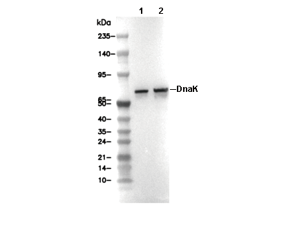 Lane 1: DnaK active recombinant protein, Lane 2: E.coli