|
Cómo citar 1. Para citas en el texto (Materiales y métodos): 2. Para la tabla de recursos clave: |
||
|
Llamada gratuita: (877) 796-6397 -- Solo EE. UU. y Canadá -- |
Fax: +1-832-582-8590 Pedidos: +1-832-582-8158 |
Soporte técnico: +1-832-582-8158 Ext:3 Por favor, indique su número de pedido en el correo electrónico. Nos esforzamos por responder a todas las consultas por correo electrónico en el plazo de un día hábil. |
Descripción biológica
| Especificidad | Daxx Antibody [H1F20] detecta los niveles endógenos de la proteína Daxx total. |
|---|---|
| Antecedentes | Daxx (Death domain-associated protein) es una proteína de andamiaje nuclear expresada ubicuamente, inicialmente identificada como un interactor de Fas, que se localiza principalmente en los cuerpos nucleares de la leucemia promielocítica (PML-NBs) y es esencial para la remodelación de la cromatina, la regulación transcripcional y la modulación de la apoptosis; la inactivación de Daxx en ratones provoca letalidad embrionaria debido a una apoptosis del desarrollo excesiva. Daxx contiene un dominio de haz helicoidal N-terminal (4HB) que media las interacciones con socios como ATRX, p53 y MDM2; una región central de unión a H3.3/H4 que se pliega con dímeros de histonas para funcionar como una chaperona específica de H3.3, depositando H3.3 en los PML-NBs y telómeros; motivos poli-Asp/Glu (polyD/E) que detectan proteínas mal plegadas y facilitan su replegamiento; dos motivos de interacción con SUMO (SIMs) que regulan la sumoilación y la focalización en los PML-NBs; y un dominio C-terminal que se asocia con Fas, CENP-C y las ADN metiltransferasas. Daxx facilita la deposición de H3.3 dependiente de ATRX para el silenciamiento de la heterocromatina y el mantenimiento de los telómeros, ensambla la holoenzima de la telomerasa en los cuerpos de Cajal a través de interacciones N-terminales con subunidades TERT (DKC1, GAR1, NHP2) para promover la focalización de la telomerasa y prevenir el acortamiento de los telómeros, reprime la transcripción de los objetivos de NF-κB y E2F-1 a través del reclutamiento de HDAC y el andamiaje de los PML-NBs, modula la apoptosis a través de la activación de Fas/JNK mientras exhibe efectos anti-apoptóticos durante el desarrollo, y responde al estrés celular uniendo proteínas mal plegadas para mantener la proteostasis. Las mutaciones de Daxx asociadas a enfermedades alteran la unión a socios, como el deterioro de la deposición mediada por H3.3/ATRX o el bloqueo del ensamblaje de la telomerasa a través de lesiones N-terminales, mientras que la sumoilación postraduccional mejora la focalización de PML, la fosforilación modula la localización subnuclear, y las interacciones con las vías HIPK2 y TGF-β integran señales para el control del ciclo celular y la supervivencia. La desregulación de Daxx contribuye al cáncer a través de perturbaciones de H3.3 que promueven la expresión de oncogenes, a los síndromes de alfa-talasemia a través de mutaciones de ATRX, y a las enfermedades neurodegenerativas debido a fallos en la proteostasis. |
Información de uso
| Aplicación | WB, IF | Dilución |
|
||||
|---|---|---|---|---|---|---|---|
| Reactividad | Human, Mouse, Rat | ||||||
| Fuente | Rabbit Monoclonal Antibody | MW | 110 kDa | ||||
| Tampón de almacenamiento | PBS, pH 7.2+50% Glycerol+0.05% BSA+0.01% NaN3 | Almacenamiento (Desde la fecha de recepción) |
-20°C (avoid freeze-thaw cycles), 2 years | ||||
| WB |
Experimental Protocol:
Sample preparation
1. Tissue: Lyse the tissue sample by adding an appropriate volume of ice-cold RIPA/NP-40 Lysis Buffer (containing Protease Inhibitor Cocktail),and homogenize the tissue at a low temperature. 2. Adherent cell: Aspirate the culture medium and wash the cells with ice-cold PBS twice. Lyse the cells by adding an appropriate volume of RIPA/NP-40 Lysis Buffer (containing Protease Inhibitor Cocktail) and put the sample on ice for 5 min. 3. Suspension cell: Transfer the culture medium to a pre-cooled centrifuge tube. Centrifuge and aspirate the supernatant. Wash the cells with ice-cold PBS twice. Lyse the cells by adding an appropriate volume of RIPA/NP-40 Lysis Buffer (containing Protease Inhibitor Cocktail) and put the sample on ice for 5 min. 4. Place the lysate into a pre-cooled microcentrifuge tube. Centrifuge at 4°C for 15 min. Collect the supernatant;
5. Remove a small volume of lysate to determine the protein concentration;
6. Combine the lysate with protein loading buffer. Boil 20 µL sample under 95-100°C for 5 min. Centrifuge for 5 min after cool down on ice.
Electrophoretic separation
1. According to the concentration of extracted protein, load appropriate amount of protein sample and marker onto SDS-PAGE gels for electrophoresis. Recommended separating gel (lower gel) concentration: 5%. Reference Table for Selecting SDS-PAGE Separation Gel Concentrations 2. Power up 80V for 30 minutes. Then the power supply is adjusted (110 V~150 V), the Marker is observed, and the electrophoresis can be stopped when the indicator band of the predyed protein Marker where the protein is located is properly separated. (Note that the current should not be too large when electrophoresis, too large current (more than 150 mA) will cause the temperature to rise, affecting the result of running glue. If high currents cannot be avoided, an ice bath can be used to cool the bath.)
Transfer membrane
1. Take out the converter, soak the clip and consumables in the pre-cooled converter;
2. Activate PVDF membrane with methanol for 1 min and rinse with transfer buffer;
3. Install it in the order of "black edge of clip - sponge - filter paper - filter paper - glue -PVDF membrane - filter paper - filter paper - sponge - white edge of clip"; 4. The protein was electrotransferred to PVDF membrane. ( 0.45 µm PVDF membrane is recommended ) Reference Table for Selecting PVDF Membrane Pore Size Specifications Recommended conditions for wet transfer: 200 mA, 120 min. ( Note that the transfer conditions can be adjusted according to the protein size. For high-molecular-weight proteins, a higher current and longer transfer time are recommended. However, ensure that the transfer tank remains at a low temperature to prevent gel melting.)
Block
1. After electrotransfer, wash the film with TBST at room temperature for 5 minutes;
2. Incubate the film in the blocking solution for 1 hour at room temperature;
3. Wash the film with TBST for 3 times, 5 minutes each time.
Antibody incubation
1. Use 5% skim milk powder to prepare the primary antibody working liquid (recommended dilution ratio for primary antibody 1:1000), gently shake and incubate with the film at 4°C overnight; 2. Wash the film with TBST 3 times, 5 minutes each time;
3. Add the secondary antibody to the blocking solution and incubate with the film gently at room temperature for 1 hour;
4. After incubation, wash the film with TBST 3 times for 5 minutes each time.
Antibody staining
1. Add the prepared ECL luminescent substrate (or select other color developing substrate according to the second antibody) and mix evenly;
2. Incubate with the film for 1 minute, remove excess substrate (keep the film moist), wrap with plastic film, and expose in the imaging system.
|
| IF |
Experimental Protocol:
Sample Preparation
1. Adherent Cells: Place a clean, sterile coverslip in a culture dish. Once the cells grow to near confluence as a monolayer, remove the coverslip for further use.
2. Suspension Cells: Seed the cells onto a clean, sterile slide coated with poly-L-lysine.
3. Frozen Sections: Allow the slide to thaw at room temperature. Wash it with pure water or PBS for 2 times, 3 minutes each time.
4. Paraffin Sections: Deparaffinization and rehydration. Wash the slide with pure water or PBS for 3 times, 3 minutes each time. Then perform antigen retrieval.
Fixation
1. Fix the cell coverslips/spots or tissue sections at room temperature using a fixative such as 4% paraformaldehyde (4% PFA) for 10-15 minutes.
2. Wash the sample with PBS for 3 times, 3 minutes each time.
Permeabilization
1.Add a detergent such as 0.1–0.3% Triton X-100 to the sample and incubate at room temperature for 10–20 minutes.
(Note: This step is only required for intracellular antigens. For antigens expressed on the cell membrane, this step is unnecessary.)
Wash the sample with PBS for 3 times, 3 minutes each time.
Blocking
Add blocking solution and incubate at room temperature for at least 1 hour. (Common blocking solutions include: serum from the same source as the secondary antibody, BSA, or goat serum.)
Note: Ensure the sample remains moist during and after the blocking step to prevent drying, which can lead to high background.
Immunofluorescence Staining (Day 1)
1. Remove the blocking solution and add the diluted primary antibody.
2. Incubate the sample in a humidified chamber at 4°C overnight.
Immunofluorescence Staining (Day 2)
1. Remove the primary antibody and wash with PBST for 3 times, 5 minutes each time.
2. Add the diluted fluorescent secondary antibody and incubate in the dark at 4°C for 1–2 hours.
3. Remove the secondary antibody and wash with PBST for 3 times, 5 minutes each time.
4. Add diluted DAPI and incubate at room temperature in the dark for 5–10 minutes.
5. Wash with PBST for 3 times, 5 minutes each time.
Mounting
1. Mount the sample with an anti-fade mounting medium.
2. Allow the slide to dry at room temperature overnight in the dark.
3. Store the slide in a slide storage box at 4°C, protected from light.
|
Referencias
|
Datos de aplicación
WB
Validado por Selleck
-
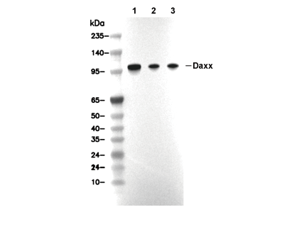 Lane 1: K562, Lane 2: A20, Lane 3: PC12