|
Cómo citar 1. Para citas en el texto (Materiales y métodos): 2. Para la tabla de recursos clave: |
||
|
Llamada gratuita: (877) 796-6397 -- Solo EE. UU. y Canadá -- |
Fax: +1-832-582-8590 Pedidos: +1-832-582-8158 |
Soporte técnico: +1-832-582-8158 Ext:3 Por favor, indique su número de pedido en el correo electrónico. Nos esforzamos por responder a todas las consultas por correo electrónico en el plazo de un día hábil. |
Descripción biológica
| Especificidad | Collagen IV Antibody [E5G18] detecta niveles endógenos de proteína Collagen IV total. |
|---|---|
| Antecedentes | El Collagen IV es el colágeno estructural principal de las membranas basales, matrices extracelulares especializadas que proporcionan soporte mecánico, regulan la adhesión celular y organizan otros componentes de la matriz. Es un heterotrímero compuesto por combinaciones de seis cadenas α (α1–α6) codificadas por genes distintos, típicamente dispuestas como dos cadenas α1 y una cadena α2 en la mayoría de los tejidos, ensambladas intracelularmente en protómeros de triple hélice. Estos protómeros se secretan y se reticulan a través de sus dominios NC1 y 7S para formar una red estable, similar a una lámina, que une lamininas, proteoglicanos y factores de crecimiento. El Collagen IV se expresa ampliamente en las membranas basales epiteliales y endoteliales, con una composición de isoformas específica de tejido, y desempeña roles esenciales en el desarrollo, la integridad tisular, la migración celular y la señalización a través de Integrin como α1β1 y α2β1. Los defectos o la reticulación aberrante del colágeno IV contribuyen a diversas patologías, incluyendo el síndrome de Alport, la enfermedad de Goodpasture, la nefropatía diabética y ciertos trastornos dermatológicos. |
Información de uso
| Aplicación | IHC | Dilución |
|
||
|---|---|---|---|---|---|
| Reactividad | Human | ||||
| Fuente | Mouse Monoclonal Antibody | MW | |||
| Tampón de almacenamiento | PBS, pH 7.2+50% Glycerol+0.05% BSA+0.01% NaN3 | Almacenamiento (Desde la fecha de recepción) |
-20°C (avoid freeze-thaw cycles), 2 years | ||
| IHC |
Experimental Protocol:
Deparaffinization/Rehydration
1. Deparaffinize/hydrate sections:
2. Incubate sections in three washes of xylene for 5 min each.
3. Incubate sections in two washes of 100% ethanol for 10 min each.
4. Incubate sections in two washes of 95% ethanol for 10 min each.
5. Wash sections two times in dH2O for 5 min each.
6.Antigen retrieval: For Citrate: Heat slides in a microwave submersed in 1X citrate unmasking solution until boiling is initiated; continue with 10 min at a sub-boiling temperature (95°-98°C). Cool slides on bench top for 30 min.
Staining
1. Wash sections in dH2O three times for 5 min each.
2. Incubate sections in 3% hydrogen peroxide for 10 min.
3. Wash sections in dH2O two times for 5 min each.
4. Wash sections in wash buffer for 5 min.
5. Block each section with 100–400 µl of blocking solution for 1 hr at room temperature.
6. Remove blocking solution and add 100–400 µl primary antibody diluent in to each section. Incubate overnight at 4°C.
7. Remove antibody solution and wash sections with wash buffer three times for 5 min each.
8. Cover section with 1–3 drops HRPas needed. Incubate in a humidified chamber for 30 min at room temperature.
9. Wash sections three times with wash buffer for 5 min each.
10. Add DAB Chromogen Concentrate to DAB Diluent and mix well before use.
11. Apply 100–400 µl DAB to each section and monitor closely. 1–10 min generally provides an acceptable staining intensity.
12. Immerse slides in dH2O.
13. If desired, counterstain sections with hematoxylin.
14. Wash sections in dH2O two times for 5 min each.
15. Dehydrate sections: Incubate sections in 95% ethanol two times for 10 sec each; Repeat in 100% ethanol, incubating sections two times for 10 sec each; Repeat in xylene, incubating sections two times for 10 sec each.
16. Mount sections with coverslips and mounting medium.
|
Referencias
|
Datos de aplicación
IHC
Validado por Selleck
-
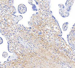 Immunohistochemical analysis of formalin fixed paraffin embedded human placenta tissue with F1667 at 1:125 dilution.