|
Cómo citar 1. Para citas en el texto (Materiales y métodos): 2. Para la tabla de recursos clave: |
||
|
Llamada gratuita: (877) 796-6397 -- Solo EE. UU. y Canadá -- |
Fax: +1-832-582-8590 Pedidos: +1-832-582-8158 |
Soporte técnico: +1-832-582-8158 Ext:3 Por favor, indique su número de pedido en el correo electrónico. Nos esforzamos por responder a todas las consultas por correo electrónico en el plazo de un día hábil. |
Descripción biológica
| Especificidad | BRAF Antibody [L21P11] detecta los niveles endógenos de la proteína BRAF total. |
|---|---|
| Antecedentes | El gen BRAF, localizado en el cromosoma 7q34, codifica una proteína quinasa serina/treonina que es un componente central de la vía de señalización de la MAP quinasa/ERK. Esta vía rige procesos celulares clave, incluyendo el crecimiento, la proliferación, la diferenciación, la senescencia y la apoptosis. Dentro de la cascada Ras/Raf/MEK/ERK, BRAF funciona como un efector directo descendente de RAS, activándose a través de la dimerización. Una vez activado, BRAF fosforila y activa MEK, que posteriormente fosforila ERK. La ERK activada regula luego proteínas citoplasmáticas y factores de transcripción que mejoran la supervivencia, proliferación y motilidad celular mientras suprimen la diferenciación. Las mutaciones en BRAF interrumpen la unión regulada entre RAS, BRAF y MEK, lo que lleva a la activación espontánea de la cascada y a una señalización persistente a través de MEK y ERK. Dicha desregulación juega un papel importante en la oncogénesis y en los síndromes del desarrollo. Las mutaciones de BRAF se detectan en aproximadamente el 7% de todos los cánceres humanos, con una prevalencia particularmente alta en ciertos tipos de tumores: casi el 100% de las leucemias de células pilosas (HCL), 50-60% de los melanomas, 30-50% de los carcinomas papilares de tiroides, 10-20% de los cánceres colorrectales y 3-5% de los cánceres de pulmón no microcíticos. La mutación más frecuente implica una sustitución en el codón 600, que normalmente codifica valina (V600). En hasta el 98% de los casos mutados de BRAF, este residuo es reemplazado por ácido glutámico (V600E, la variante predominante), lisina (V600K, presente en el 5-10% de los casos), o menos comúnmente por otros aminoácidos como alanina, ácido aspártico, glicina, leucina, metionina, glutamina o arginina (V600A/D/G/L/M/Q/R, que en conjunto representan hasta el 5%). Estas mutaciones hacen que BRAF sea constitutivamente activo como monómero, evitando el requisito de activación mediada por RAS e impulsando una señalización incontrolada a través de la vía MAPK. |
Información de uso
| Aplicación | WB, IP, IHC | Dilución |
|
||||||
|---|---|---|---|---|---|---|---|---|---|
| Reactividad | Mouse, Rat, Human | ||||||||
| Fuente | Rabbit Monoclonal Antibody | MW | 85 kDa | ||||||
| Tampón de almacenamiento | PBS, pH 7.2+50% Glycerol+0.05% BSA+0.01% NaN3 | Almacenamiento (Desde la fecha de recepción) |
-20°C (avoid freeze-thaw cycles), 2 years | ||||||
| IHC |
Experimental Protocol:
Deparaffinization/Rehydration
1. Deparaffinize/hydrate sections:
2. Incubate sections in three washes of xylene for 5 min each.
3. Incubate sections in two washes of 100% ethanol for 10 min each.
4. Incubate sections in two washes of 95% ethanol for 10 min each.
5. Wash sections two times in dH2O for 5 min each.
6.Antigen retrieval: For Citrate: Heat slides in a microwave submersed in 1X citrate unmasking solution until boiling is initiated; continue with 10 min at a sub-boiling temperature (95°-98°C). Cool slides on bench top for 30 min.
Staining
1. Wash sections in dH2O three times for 5 min each.
2. Incubate sections in 3% hydrogen peroxide for 10 min.
3. Wash sections in dH2O two times for 5 min each.
4. Wash sections in wash buffer for 5 min.
5. Block each section with 100–400 µl of blocking solution for 1 hr at room temperature.
6. Remove blocking solution and add 100–400 µl primary antibody diluent in to each section. Incubate overnight at 4°C.
7. Remove antibody solution and wash sections with wash buffer three times for 5 min each.
8. Cover section with 1–3 drops HRPas needed. Incubate in a humidified chamber for 30 min at room temperature.
9. Wash sections three times with wash buffer for 5 min each.
10. Add DAB Chromogen Concentrate to DAB Diluent and mix well before use.
11. Apply 100–400 µl DAB to each section and monitor closely. 1–10 min generally provides an acceptable staining intensity.
12. Immerse slides in dH2O.
13. If desired, counterstain sections with hematoxylin.
14. Wash sections in dH2O two times for 5 min each.
15. Dehydrate sections: Incubate sections in 95% ethanol two times for 10 sec each; Repeat in 100% ethanol, incubating sections two times for 10 sec each; Repeat in xylene, incubating sections two times for 10 sec each.
16. Mount sections with coverslips and mounting medium.
|
| WB |
Experimental Protocol:
Sample preparation
1. Tissue: Lyse the tissue sample by adding an appropriate volume of ice-cold RIPA/NP-40 Lysis Buffer (containing Protease Inhibitor Cocktail),and homogenize the tissue at a low temperature. 2. Adherent cell: Aspirate the culture medium and wash the cells with ice-cold PBS twice. Lyse the cells by adding an appropriate volume of RIPA/NP-40 Lysis Buffer (containing Protease Inhibitor Cocktail) and put the sample on ice for 5 min. 3. Suspension cell: Transfer the culture medium to a pre-cooled centrifuge tube. Centrifuge and aspirate the supernatant. Wash the cells with ice-cold PBS twice. Lyse the cells by adding an appropriate volume of RIPA/NP-40 Lysis Buffer (containing Protease Inhibitor Cocktail) and put the sample on ice for 5 min. 4. Place the lysate into a pre-cooled microcentrifuge tube. Centrifuge at 4°C for 15 min. Collect the supernatant;
5. Remove a small volume of lysate to determine the protein concentration;
6. Combine the lysate with protein loading buffer. Boil 20 µL sample under 95-100°C for 5 min. Centrifuge for 5 min after cool down on ice.
Electrophoretic separation
1. According to the concentration of extracted protein, load appropriate amount of protein sample and marker onto SDS-PAGE gels for electrophoresis. Recommended separating gel (lower gel) concentration: 10%. Reference Table for Selecting SDS-PAGE Separation Gel Concentrations 2. Power up 80V for 30 minutes. Then the power supply is adjusted (110 V~150 V), the Marker is observed, and the electrophoresis can be stopped when the indicator band of the predyed protein Marker where the protein is located is properly separated. (Note that the current should not be too large when electrophoresis, too large current (more than 150 mA) will cause the temperature to rise, affecting the result of running glue. If high currents cannot be avoided, an ice bath can be used to cool the bath.)
Transfer membrane
1. Take out the converter, soak the clip and consumables in the pre-cooled converter;
2. Activate PVDF membrane with methanol for 1 min and rinse with transfer buffer;
3. Install it in the order of "black edge of clip - sponge - filter paper - filter paper - glue -PVDF membrane - filter paper - filter paper - sponge - white edge of clip"; 4. The protein was electrotransferred to PVDF membrane. ( 0.45 µm PVDF membrane is recommended ) Reference Table for Selecting PVDF Membrane Pore Size Specifications Recommended conditions for wet transfer: 200 mA, 120 min. ( Note that the transfer conditions can be adjusted according to the protein size. For high-molecular-weight proteins, a higher current and longer transfer time are recommended. However, ensure that the transfer tank remains at a low temperature to prevent gel melting.)
Block
1. After electrotransfer, wash the film with TBST at room temperature for 5 minutes;
2. Incubate the film in the blocking solution for 1 hour at room temperature;
3. Wash the film with TBST for 3 times, 5 minutes each time.
Antibody incubation
1. Use 5% skim milk powder to prepare the primary antibody working liquid (recommended dilution ratio for primary antibody 1:1000), gently shake and incubate with the film at 4°C overnight; 2. Wash the film with TBST 3 times, 5 minutes each time;
3. Add the secondary antibody to the blocking solution and incubate with the film gently at room temperature for 1 hour;
4. After incubation, wash the film with TBST 3 times for 5 minutes each time.
Antibody staining
1. Add the prepared ECL luminescent substrate (or select other color developing substrate according to the second antibody) and mix evenly;
2. Incubate with the film for 1 minute, remove excess substrate (keep the film moist), wrap with plastic film, and expose in the imaging system.
|
Referencias
|
Datos de aplicación
WB
Validado por Selleck
-
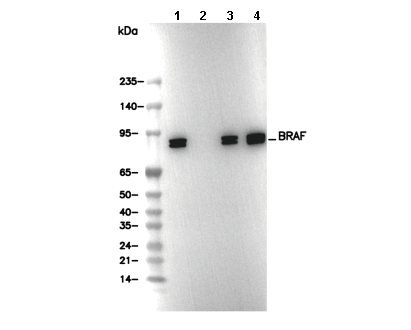 Lane 1: HeLa, Lane 2: HeLa (KO BRAF), Lane 3: HeLa, Lane 4: Mouse brain