|
Cómo citar 1. Para citas en el texto (Materiales y métodos): 2. Para la tabla de recursos clave: |
||
|
Llamada gratuita: (877) 796-6397 -- Solo EE. UU. y Canadá -- |
Fax: +1-832-582-8590 Pedidos: +1-832-582-8158 |
Soporte técnico: +1-832-582-8158 Ext:3 Por favor, indique su número de pedido en el correo electrónico. Nos esforzamos por responder a todas las consultas por correo electrónico en el plazo de un día hábil. |
Descripción biológica
| Especificidad | ATG4B Antibody [P14M3] detecta niveles endógenos de proteína ATG4B total. |
|---|---|
| Antecedentes | ATG4B es una proteasa de cisteína crucial en la Autophagy, responsable del procesamiento y reciclaje de proteínas de la familia ATG8 como LC3 y GABARAP, lo que permite la biogénesis del autofagosoma en eucariotas. ATG4B contiene una tríada catalítica compuesta por Cys74, Asp278 e His280 dentro de un pliegue similar a la papaína, con un bucle regulador y Trp142 que mantienen la enzima en un estado autoinhibido hasta que el compromiso del sustrato induce cambios conformacionales para la hidrólisis del enlace peptídico. Los motivos N-terminales mejoran aún más la especificidad para LC3 conjugado a fosfatidiletanolamina (PE) en las membranas autofágicas. La función principal de ATG4B es exponer la glicina C-terminal en pro-LC3 mediante una escisión de cebado inicial, preparándola para la conjugación a PE a través de la cascada similar a ubiquitina ATG7/ATG3, y posteriormente catalizar la deslipidación de LC3-PE en la membrana autofagosómica externa, facilitando el reciclaje de LC3 libre. Con la selectividad de sustrato más amplia entre los parálogos de ATG4, ATG4B favorece principalmente a LC3, equilibrando la elongación de la membrana del autofagosoma, el anclaje y el flujo autofágico. Su actividad está estrictamente regulada a través de modificaciones redox en Cys292 y Cys361 y por fosforilación, adaptando la Autophagy al estrés celular; la sobreexpresión de ATG4B de tipo salvaje puede bloquear la lipidación de LC3 por desconjugación excesiva, mientras que mutantes catalíticamente inactivos como C74A secuestran LC3 e inhiben la Autophagy. La desregulación de la actividad de ATG4B está relacionada con la tumorigénesis debido a un flujo autofágico alterado. |
Información de uso
| Aplicación | WB, FCM | Dilución |
|
||||
|---|---|---|---|---|---|---|---|
| Reactividad | Mouse, Human | ||||||
| Fuente | Rabbit Monoclonal Antibody | MW | 44 kDa | ||||
| Tampón de almacenamiento | PBS, pH 7.2+50% Glycerol+0.05% BSA+0.01% NaN3 | Almacenamiento (Desde la fecha de recepción) |
-20°C (avoid freeze-thaw cycles), 2 years | ||||
| WB |
Experimental Protocol:
Sample preparation
1. Tissue: Lyse the tissue sample by adding an appropriate volume of ice-cold RIPA/NP-40 Lysis Buffer (containing Protease Inhibitor Cocktail),and homogenize the tissue at a low temperature. 2. Adherent cell: Aspirate the culture medium and wash the cells with ice-cold PBS twice. Lyse the cells by adding an appropriate volume of RIPA/NP-40 Lysis Buffer (containing Protease Inhibitor Cocktail) and put the sample on ice for 5 min. 3. Suspension cell: Transfer the culture medium to a pre-cooled centrifuge tube. Centrifuge and aspirate the supernatant. Wash the cells with ice-cold PBS twice. Lyse the cells by adding an appropriate volume of RIPA/NP-40 Lysis Buffer (containing Protease Inhibitor Cocktail) and put the sample on ice for 5 min. 4. Place the lysate into a pre-cooled microcentrifuge tube. Centrifuge at 4°C for 15 min. Collect the supernatant;
5. Remove a small volume of lysate to determine the protein concentration;
6. Combine the lysate with protein loading buffer. Boil 20 µL sample under 95-100°C for 5 min. Centrifuge for 5 min after cool down on ice.
Electrophoretic separation
1. According to the concentration of extracted protein, load appropriate amount of protein sample and marker onto SDS-PAGE gels for electrophoresis. Recommended separating gel (lower gel) concentration: 10%. Reference Table for Selecting SDS-PAGE Separation Gel Concentrations 2. Power up 80V for 30 minutes. Then the power supply is adjusted (110 V~150 V), the Marker is observed, and the electrophoresis can be stopped when the indicator band of the predyed protein Marker where the protein is located is properly separated. (Note that the current should not be too large when electrophoresis, too large current (more than 150 mA) will cause the temperature to rise, affecting the result of running glue. If high currents cannot be avoided, an ice bath can be used to cool the bath.)
Transfer membrane
1. Take out the converter, soak the clip and consumables in the pre-cooled converter;
2. Activate PVDF membrane with methanol for 1 min and rinse with transfer buffer;
3. Install it in the order of "black edge of clip - sponge - filter paper - filter paper - glue -PVDF membrane - filter paper - filter paper - sponge - white edge of clip"; 4. The protein was electrotransferred to PVDF membrane. ( 0.45 µm PVDF membrane is recommended ) Reference Table for Selecting PVDF Membrane Pore Size Specifications Recommended conditions for wet transfer: 200 mA, 120 min. ( Note that the transfer conditions can be adjusted according to the protein size. For high-molecular-weight proteins, a higher current and longer transfer time are recommended. However, ensure that the transfer tank remains at a low temperature to prevent gel melting.)
Block
1. After electrotransfer, wash the film with TBST at room temperature for 5 minutes;
2. Incubate the film in the blocking solution for 1 hour at room temperature;
3. Wash the film with TBST for 3 times, 5 minutes each time.
Antibody incubation
1. Use 5% skim milk powder to prepare the primary antibody working liquid (recommended dilution ratio for primary antibody 1:1000), gently shake and incubate with the film at 4°C overnight; 2. Wash the film with TBST 3 times, 5 minutes each time;
3. Add the secondary antibody to the blocking solution and incubate with the film gently at room temperature for 1 hour;
4. After incubation, wash the film with TBST 3 times for 5 minutes each time.
Antibody staining
1. Add the prepared ECL luminescent substrate (or select other color developing substrate according to the second antibody) and mix evenly;
2. Incubate with the film for 1 minute, remove excess substrate (keep the film moist), wrap with plastic film, and expose in the imaging system.
|
Referencias
|
Datos de aplicación
WB
Validado por Selleck
-
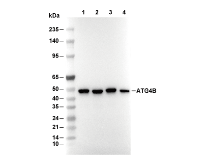 Lane 1: Ramos, Lane 2: 293T, Lane 3: Jurkat, Lane 4: Hela