Datos técnicos
| Fórmula | C32H34Cl2N4O4S |
||||||
| Peso molecular | 641.61 | Número CAS | 477575-56-7 | ||||
| Solubilidad (25°C)* | In vitro | DMSO | 128 mg/mL (199.49 mM) | ||||
| Water | Insoluble | ||||||
| Ethanol | Insoluble | ||||||
| In vivo (Agregue los solventes al producto individualmente y en orden.) |
|
||||||
|
* <1 mg/ml significa ligeramente soluble o insoluble. * Tenga en cuenta que Selleck prueba la solubilidad de todos los compuestos internamente, y la solubilidad real puede diferir ligeramente de los valores publicados. Esto es normal y se debe a ligeras variaciones entre lotes. * Envío a temperatura ambiente (Las pruebas de estabilidad demuestran que este producto se puede enviar sin medidas de refrigeración.) |
|||||||
Preparación de soluciones madre
Actividad biológica
| Descripción | PHA-665752 es un inhibidor potente, selectivo y ATP-competitivo de c-Met con una IC50 de 9 nM en ensayos libres de células, >50 veces más selectivo para c-Met que para RTKs o STKs. | ||||||
|---|---|---|---|---|---|---|---|
| Objetivos |
|
||||||
| In vitro | PHA-665752 inhibe significativamente la actividad de la quinasa c-Met con una Ki de 4 nM, y exhibe una selectividad >50 veces mayor para c-Met en comparación con diversas tirosina quinasas y serina-treonina quinasas. Este compuesto inhibe potentemente la autofosforilación de c-Met estimulada por HGF con una IC50 de 25-50 nM. También bloquea significativamente funciones dependientes de HGF y c-Met, como la motilidad celular y la proliferación celular, con IC50 de 40-50 nM y 18-42 nM, respectivamente. Además, este químico inhibe potentemente la fosforilación constitutiva o estimulada por HGF de mediadores de la vía descendente de c-Met como Gab-1, ERK, Akt, STAT3, PLC-γ y FAK en múltiples líneas celulares tumorales. Inhibe el crecimiento celular en células BaF3 transformadas por TPR-MET con una IC50 <60 nM, e inhibe la motilidad y migración celular constitutiva en un 92,5% a 0,2 μM. La inhibición de c-Met por este compuesto (0,2 μM) también induce la apoptosis celular en un 33,1% y la detención del ciclo celular en G1, con un aumento de las células en fase G1 del 42,4% al 77,0%. Puede cooperar con rapamicina para inhibir el crecimiento de células BaF3 transformadas por TPR-MET y células H441 de cáncer de pulmón de células no pequeñas. | ||||||
| In vivo | La administración de PHA-665752 induce una inhibición del crecimiento tumoral dependiente de la dosis en xenoinjertos S114 en un 20%, 39% y 68%, a dosis de 7,5, 15 y 30 mg/kg/día, respectivamente. Este tratamiento con el compuesto reduce significativamente el crecimiento tumoral de NCI-H69, NCI-H441 y A549 en xenoinjertos de ratón en un 99%, 75% y 59%, respectivamente. También inhibe significativamente la angiogénesis en más del 85%, debido a la disminución de la producción del factor de crecimiento endotelial vascular y al aumento de la producción del inhibidor de la angiogénesis trombospondina-1. |
Protocolo (de referencia)
| Ensayo de quinasa:[1] |
|
|---|---|
| Ensayo celular:[1] |
|
| Estudio en animales:[1] |
|
Referencias
|
Validación de productos por parte del cliente
-
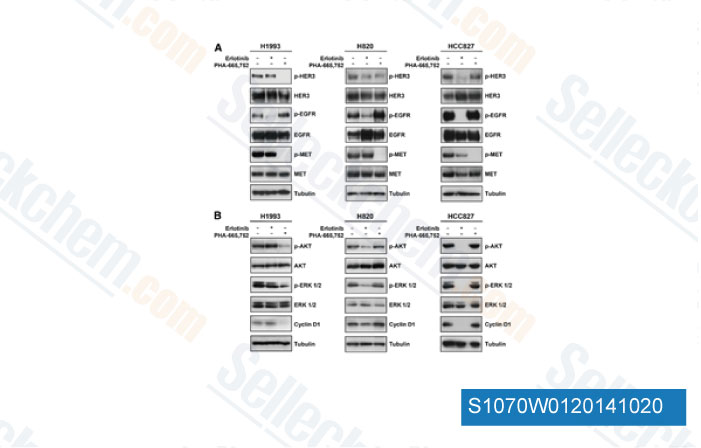
Datos de [ Clin Cancer Res , 2014 , 20, 4806-15 ]
-
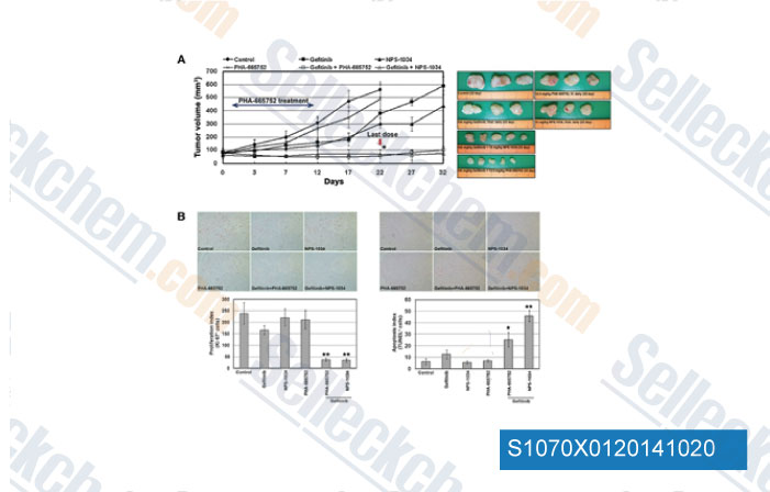
Datos de [ Cancer Res , 2014 , 74, 253-62 ]
-
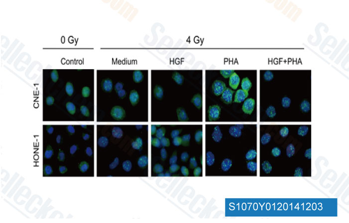
Datos de [ Biochem Biophys Res Commun , 2014 , 449(1), 49-54 ]
-
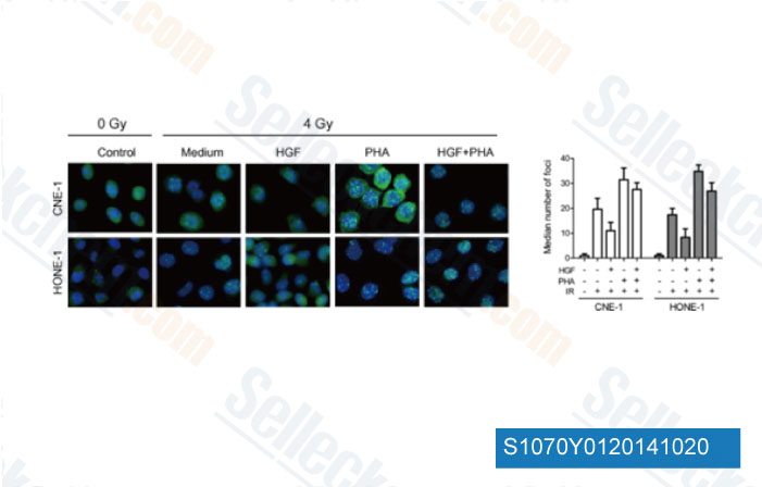
Datos de [ Biochem Biophys Res Commun , 2014 , 449, 49-54 ]
Sellecks PHA-665752 Ha sido citado por 100 Publicaciones
| Inhibition of TFF3 synergizes with c-MET inhibitors to decrease the CSC-like phenotype and metastatic burden in ER+HER2+ mammary carcinoma [ Cell Death Dis, 2025, 16(1):76] | PubMed: 39920140 |
| Macrophage Subpopulation Promotes Skeletal Muscle Regeneration Through HGF/MET Signaling-Mediated Skeletal Muscle Stem Cell Proliferation [ Aging Cell, 2025, e70042.] | PubMed: 40132988 |
| Overcoming MET-targeted drug resistance in MET-amplified lung cancer by aurora kinase B inhibition [ Biochim Biophys Acta Mol Cell Res, 2025, 1872(7):120001] | PubMed: 40499687 |
| MET receptor serves as a promising target in melanoma brain metastases [ Acta Neuropathol, 2024, 147(1):44] | PubMed: 38386085 |
| LncRNA CHROMR/miR-27b-3p/MET axis promotes the proliferation, invasion, and contributes to rituximab resistance in diffuse large B-cell lymphoma [ J Biol Chem, 2024, 300(3):105762] | PubMed: 38367665 |
| Single targeting of MET in EGFR-mutated and MET-amplified non-small cell lung cancer [ Br J Cancer, 2023, 128(12):2186-2196] | PubMed: 37059804 |
| Cancer-Associated Fibroblasts Promote Radioresistance of Breast Cancer Cells via the HGF/c-Met Signaling Pathway [ Int J Radiat Oncol Biol Phys, 2023, 116(3):640-654] | PubMed: 36586496 |
| MET Receptor Tyrosine Kinase Inhibition Reduces Interferon-Gamma (IFN-γ)-Stimulated PD-L1 Expression through the STAT3 Pathway in Melanoma Cells [ Cancers (Basel), 2023, 15(13)3408] | PubMed: 37444518 |
| Cabozantinib inhibits HBV-RNA transcription by decreasing STAT3 binding to the enhancer region of cccDNA [ Hepatol Commun, 2023, 10.1097/HC9.0000000000000313] | PubMed: 37938099 |
| Dual-targeting therapy against HER3/MET in human colorectal cancers [ Cancer Med, 2023, 10.1002/cam4.5673] | PubMed: 36751113 |
POLÍTICA DE DEVOLUCIÓN
La Política de Devolución Incondicional de Selleck Chemical garantiza una experiencia de compra en línea fluida para nuestros clientes. Si no está satisfecho con su compra de alguna manera, puede devolver cualquier artículo(s) dentro de los 7 días posteriores a su recepción. En caso de problemas de calidad del producto, ya sean problemas relacionados con el protocolo o con el producto, puede devolver cualquier artículo(s) dentro de los 365 días a partir de la fecha de compra original. Siga las instrucciones a continuación al devolver productos.
ENVÍO Y ALMACENAMIENTO
Los productos Selleck se transportan a temperatura ambiente. Si recibe el producto a temperatura ambiente, tenga la seguridad de que el Departamento de Inspección de Calidad de Selleck ha realizado experimentos para verificar que la colocación a temperatura normal durante un mes no afectará la actividad biológica de los productos en polvo. Después de la recogida, guarde el producto de acuerdo con los requisitos descritos en la hoja de datos. La mayoría de los productos Selleck son estables en las condiciones recomendadas.
NO PARA USO HUMANO, DIAGNÓSTICO VETERINARIO O TERAPÉUTICO.