OSI-930

N.º de catálogoS1220 Lote:S122002

Imprimir

Datos técnicos

Fórmula

C22H16F3N3O2S

Peso molecular 443.44 Número CAS 728033-96-3
Solubilidad (25°C)* In vitro DMSO 89 mg/mL (200.7 mM)
Ethanol 3 mg/mL (6.76 mM)
Water Insoluble
In vivo (Agregue los solventes al producto individualmente y en orden.)
Homogeneous suspension
CMC-NA
≥5mg/ml Taking the 1 mL working solution as an example, add 5 mg of this product to 1 ml of CMC-Na solution, mix evenly to obtain a homogeneous suspension with a final concentration of 5 mg/ml.
Clear solution
30%PEG400 0.5%Tween80 5%propylene glycol

Validado por los laboratorios Selleck. Si necesita ajustes en esta formulación, póngase en contacto con nuestro equipo de ventas para pruebas personalizadas.

5.000mg/ml (11.28mM) Taking the 1 mL working solution as an example, add 300 μL of 16.67 mg/ml clarified PEG400 stock solution to 5 μL of Tween80, mix evenly to clarify it; add 50 μL Propylene glycol to the above system, mix evenly to clarify it; then continue to add 645 μL ddH2O Dilute to 1 mL. The mixed solution should be used immediately for optimal results. 
* <1 mg/ml significa ligeramente soluble o insoluble.
* Tenga en cuenta que Selleck prueba la solubilidad de todos los compuestos internamente, y la solubilidad real puede diferir ligeramente de los valores publicados. Esto es normal y se debe a ligeras variaciones entre lotes.
* Envío a temperatura ambiente (Las pruebas de estabilidad demuestran que este producto se puede enviar sin medidas de refrigeración.)

Preparación de soluciones madre

Actividad biológica

Descripción OSI-930 es un potente inhibidor de Kit (c-Kit), KDR y CSF-1R con IC50 de 80 nM, 9 nM y 15 nM, respectivamente; este compuesto también es potente para Flt-1, c-Raf y Lck y tiene baja actividad contra PDGFRα/β, Flt-3 y Abl. Fase 1.
Objetivos
FLT1
(Cell-free assay)
KDR
(Cell-free assay)
CSF-1R
(Cell-free assay)
LCK
(Cell-free assay)
C-Raf
(Cell-free assay)
Ver más
8 nM 9 nM 15 nM 22 nM 41 nM
In vitro OSI-930 inhibe la proliferación celular en la línea celular HMC-1 con una IC50 de 14 nM sin un efecto significativo en el crecimiento de la línea celular COLO-205 que no expresa un receptor de tirosina quinasa mutante constitutivamente activo. Además, este compuesto también induce la apoptosis en la línea celular HMC-1 con una EC50 de 34 nM. Un estudio reciente muestra que este químico inactiva el citocromo P450 (P450) 3A4 purificado y recombinante con un Ki de 24 μM de manera dependiente del tiempo y la concentración.
In vivo OSI-930, administrado a la dosis máxima eficaz de 200 mg/kg por sonda gástrica oral, exhibe una potente actividad antitumoral en una amplia gama de modelos de xenoinjertos preclínicos, incluyendo los modelos de xenoinjertos HMC-1, NCI-SNU-5, COLO-205 y U251.

Protocolo (de referencia)

Ensayo de quinasa:[1]
  • Ensayos de proteína quinasa

    Los ensayos de proteína quinasa se realizan internamente mediante métodos de ensayo basados en ELISA (Kit, KDR, PDGFRα y PDGFRβ) o mediante un método radiométrico. Los ensayos ELISA internos utilizaron poli(Glu:Tyr) como sustrato unido a la superficie de placas de ensayo de 96 pocillos; la fosforilación se detecta luego utilizando un anticuerpo antifosfotirosina conjugado a HRP. El anticuerpo unido se cuantifica luego usando ABTS como sustrato de peroxidasa midiendo la absorbancia a 405/490 nm. Todos los ensayos utilizan dominios catalíticos de quinasa recombinantes purificados que se expresan en células de insectos o en bacterias. La proteína Kit y EGFR utilizada para los ensayos internos se prepara internamente; otras enzimas se obtienen. La proteína Kit recombinante se expresa como una proteína de fusión glutatión S-transferasa NH2-terminal en células de insectos y se purifica inicialmente como una enzima no fosforilada (no activada) con una Km relativamente alta para ATP (400 μM). En algunos ensayos, se prepara una forma activada (tirosina fosforilada) de la enzima mediante incubación con 1 mM de ATP durante 1 hora a 30 °C. La proteína fosforilada se pasa luego a través de una columna de desalación para eliminar la mayor parte del ATP y se almacena a -80 °C en un tampón que contiene 50% de glicerol. La preparación resultante tiene una actividad específica considerablemente mayor y una Km para ATP (25 μM) menor que la preparación inicial no fosforilada. La inhibición de la autofosforilación de Kit por este compuesto se ensaya mediante incubación de la enzima no fosforilada a 30 °C en presencia de 200 μM de ATP y varias concentraciones de este químico. La reacción se detiene retirando alícuotas en tampón de muestra SDS-PAGE y luego calentando a 100 °C durante 5 minutos. El grado de fosforilación de Kit se determina luego mediante inmunoblotting para Kit total y Kit fosforilado.

Ensayo celular:[1]
  • Líneas celulares

    HMC-1 and COLO-205

  • Concentraciones

    0--1 μM

  • Tiempo de incubación

    48 hours

  • Método

    For assays of cell proliferation and apoptosis, cells are seeded into 96-well plates and incubated for 2 to 3 days in the presence of OSI-930 at various concentrations. Inhibition of cell growth is determined by luminescent quantitation of the intracellular ATP content using CellTiterGlo. Induction of caspase-dependent apoptosis by this compound is quantitated by an enzymatic caspase 3/7 assay. Inhibition of angiogenesis by this chemical is monitored using the rat aortic ring endothelial sprout outgrowth assay. Sections of aorta are prepared from CO2-euthanized male rats and cultured in vitro in a collagen matrix in the presence or absence of this compound. The collagen matrix is prepared from type 1 rat tail collagen solubilized in 0.1% acetic acid at 3 mg/mL, which is combined with 0.125 volume collagen buffer (0.05 N NaOH, 200 mM HEPES, 260 mM NaHCO3), 0.125 volume of medium 199, 0.0125 volume of 1 M NaOH, and 1% GlutaMax. Aortic rings are embedded in 0.4 mL of this matrix in six-well plates, to which 0.5 mL endothelial basal medium and the appropriate amount of this chemical is added; the rings are then incubated for 10 days and the resultant angiogenic sprout outgrowth is digitally quantitated from images by measurement of the sprout-containing area within a series of concentric rings around the aortic tissue area.

Estudio en animales:[1]
  • Modelos animales

    HMC-1, NCI-SNU-5, COLO-205 and U251 cells are injected s.c. into the right flank of CD1 nu/nu mice.

  • Dosificaciones

    ≤200 mg/kg

  • Administración

    Administered via p.o.

Referencias

  • https://pubmed.ncbi.nlm.nih.gov/16424037/
  • https://pubmed.ncbi.nlm.nih.gov/21068193/

Validación de productos por parte del cliente

RE-luc2P-HEK293 cells were pretreated with 1uM OSI-930 (green), 20uM TBB (blue), 10uM CKI-7 (purple), or 10uM H-89 (orange) for 16 h and infected with Y. enterocolitica WA or Y. pestis Ind195 at MOI 1 and 20, respectively, for 1 h. Following stimulation with 10 ng/ml TNF-α at 5 h post-infection, luciferase activity was measured 24 h post-infection. Results were determined from two independent experiments performed in triplicate. A"*" denotes that the % NF-κβ inhibition using the inhibitors was significantly different (p<0.05) compared to the no drug control (black). The relative NF-κB inhibition by Yersinia infection was determined as a percentage of luciferase activity in bacteria-infected cells relative to luciferase activity in bacteria-free control cells.

Datos de [ BMC Microbiol , 2013 , 13, 249 ]

<p> </p><p>Table 2. shows the reversal effect of OSI-930 and Fumitremorgin C (FTC) on the cytotoxicity of mitoxantrone to HEK293/pcDNA3.1 and HEK293-ABCG2-482-R2.<sup>a</sup>IC<sub>50</sub>: concentration that inhibited cell survival by 50% (means ± SD). <sup>b</sup>FR: fold-resistance was the value of that IC<sub>50</sub> value for mitoxantrone of HEK293/pcDNA3.1 cells was divided by IC<sub>50</sub> value for mitoxantrone of HEK293/pcDNA3.1 and HEK293-ABCG2-482-R2 cells in the absence or presence of OSI-930 and Fumitremorgin C (FTC). Values in table are representative of at least three independent experiments performed in triplicate.</p>

,

<p>For MTT assays, cells (2,000 ~ 5,000 cells/well) were subcultured into 96-well plates according to their growth properties. Cell proliferation was assayed at 72 hr after treatment of OSI-930 by adding 20 μl of 5 mg/ml 3-(4,5-Dimethylthiazol-2-yl)-2,5-diphenyltetrazolium bromide (MTT) solution per 100 μl of growth medium. After incubating for 3-4 h at 37°C, the media were removed and 150 µl/well of MTT solvent (either absolute DMSO or isopropanol containing 4 mM HCl and 0.1% Nonidet-40) was added to dissolve the formazan</p>

, , Dr. Yong-Weon Yi from Georgetown University Medical Center

Sellecks OSI-930 Ha sido citado por 7 Publicaciones

Orthogonal proteogenomic analysis identifies the druggable PA2G4-MYC axis in 3q26 AML [ Nat Commun, 2024, 15(1):4739] PubMed: 38834613
Small-Molecule and CRISPR Screening Converge to Reveal Receptor Tyrosine Kinase Dependencies in Pediatric Rhabdoid Tumors. [ Cell Rep, 2019, 28(9):2331-2344] PubMed: 31461650
TLR7/8-agonist-loaded Nanoparticles Promote the Polarization of Tumour-Associated Macrophages to Enhance Cancer Immunotherapy [ Nat Biomed Eng, 2018, 2(8):578-588] PubMed: 31015631
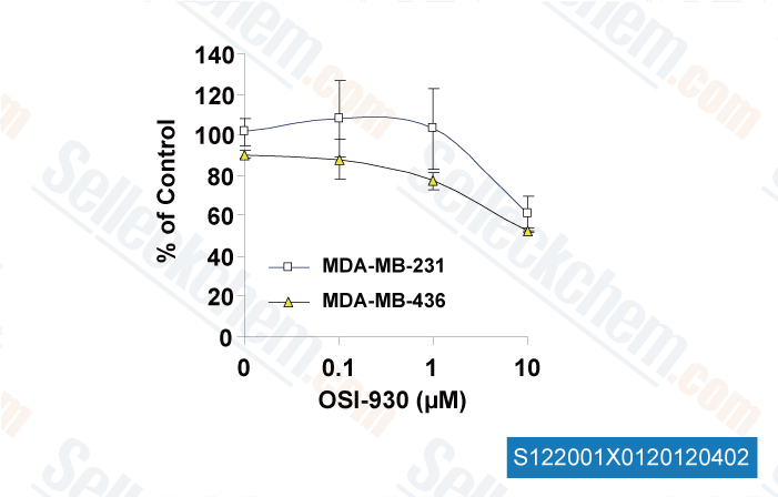
Targeting a cell state common to triple-negative breast cancers [Muellner MK, et al. Mol Syst Biol, 2015, 11(1):789] PubMed: 25699542
Dual inhibition of EGFR and MET induces synthetic lethality in triple-negative breast cancer cells through downregulation of ribosomal protein S6. [Yi YW, et al. Int J Oncol, 2015, 47(1):122-32] PubMed: 25955731
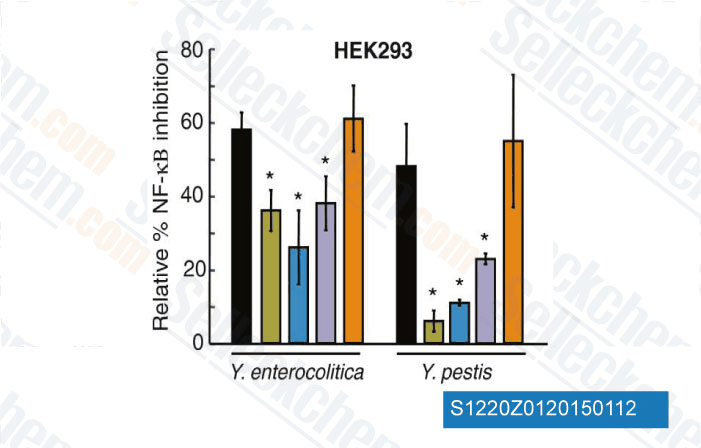
c-KIT signaling is targeted by pathogenic Yersinia to suppress the host immune response. [Micheva-Viteva SN, et al. BMC Microbiol, 2013, 13(1):249] PubMed: 24206648
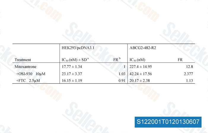
OSI-930 analogues as novel reversal agents for ABCG2-mediated multidrug resistance. [Kuang Y, et al. Biochem Pharmacol, 2012, 84(6):766-74] PubMed: 22750060

POLÍTICA DE DEVOLUCIÓN
La Política de Devolución Incondicional de Selleck Chemical garantiza una experiencia de compra en línea fluida para nuestros clientes. Si no está satisfecho con su compra de alguna manera, puede devolver cualquier artículo(s) dentro de los 7 días posteriores a su recepción. En caso de problemas de calidad del producto, ya sean problemas relacionados con el protocolo o con el producto, puede devolver cualquier artículo(s) dentro de los 365 días a partir de la fecha de compra original. Siga las instrucciones a continuación al devolver productos.

ENVÍO Y ALMACENAMIENTO
Los productos Selleck se transportan a temperatura ambiente. Si recibe el producto a temperatura ambiente, tenga la seguridad de que el Departamento de Inspección de Calidad de Selleck ha realizado experimentos para verificar que la colocación a temperatura normal durante un mes no afectará la actividad biológica de los productos en polvo. Después de la recogida, guarde el producto de acuerdo con los requisitos descritos en la hoja de datos. La mayoría de los productos Selleck son estables en las condiciones recomendadas.

NO PARA USO HUMANO, DIAGNÓSTICO VETERINARIO O TERAPÉUTICO.