Datos técnicos
| Fórmula | C29H26ClFN4O4S.2C7H8O3S |
||||||
| Peso molecular | 925.46 | Número CAS | 388082-77-7 | ||||
| Solubilidad (25°C)* | In vitro | DMSO | 100 mg/mL (108.05 mM) | ||||
| Water | Insoluble | ||||||
| Ethanol | Insoluble | ||||||
| In vivo (Agregue los solventes al producto individualmente y en orden.) |
|
||||||
|
* <1 mg/ml significa ligeramente soluble o insoluble. * Tenga en cuenta que Selleck prueba la solubilidad de todos los compuestos internamente, y la solubilidad real puede diferir ligeramente de los valores publicados. Esto es normal y se debe a ligeras variaciones entre lotes. * Envío a temperatura ambiente (Las pruebas de estabilidad demuestran que este producto se puede enviar sin medidas de refrigeración.) |
|||||||
Preparación de soluciones madre
Actividad biológica
| Descripción | Lapatinib Ditosylate es un potente inhibidor de EGFR y ErbB2 con una IC50 de 10.8 y 9.2 nM en ensayos sin células, respectivamente. | ||||||
|---|---|---|---|---|---|---|---|
| Objetivos |
|
||||||
| In vitro | Lapatinib Ditosylate inhibe débilmente la actividad de ErbB4 con una IC50 de 367 nM, y muestra una selectividad >300 veces mayor para EGFR y ErbB2 sobre otras quinasas como c-Src, c-Raf, MEK, ERK, c-Fms, CDK1, CDK2, p38, Tie-2 y VEGFR2. Este compuesto inhibe significativamente la autofosforilación del receptor de EGFR y ErbB2 de manera dosis-dependiente con una IC50 de 170 nM y 80 nM, respectivamente en células HN5; así como 210 nM y 60 nM, respectivamente en células BT474. A diferencia de OSI-774 e Iressa (ZD1839) que inhiben preferentemente el crecimiento de las células que sobreexpresan EGFR, inhibe el crecimiento de células que sobreexpresan tanto EGFR como ErbB2. Muestra una mayor actividad inhibidora contra células que sobreexpresan EGFR o ErbB2 con una IC50 de 0.09-0.21 μM, en comparación con células que expresan bajos niveles de EGFR o ErbB2 con una IC50 de 3-12 μM, y exhibe una selectividad de ~100 veces sobre las células fibroblásticas normales. Este químico inhibe potentemente el crecimiento de células HN5 y A-431 que sobreexpresan EGFR, así como de células BT474 y N87 que sobreexpresan ErbB2, e induce significativamente la detención en G1 de las células HN5 y la apoptosis de las células BT474, lo que se asocia con la inhibición de la fosforilación de AKT. | ||||||
| In vivo | La administración oral de Lapatinib Ditosylate (~100 mg/kg) dos veces al día inhibe significativamente el crecimiento de xenoinjertos BT474 y HN5 de manera dosis-dependiente. |
Protocolo (de referencia)
| Ensayo de quinasa:[1] |
|
|---|---|
| Ensayo celular:[1] |
|
| Estudio en animales:[1] |
|
Referencias
|
Validación de productos por parte del cliente
-
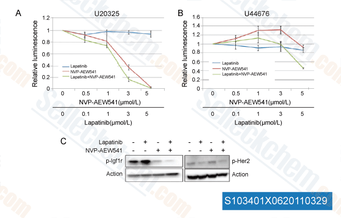
Datos de [ Mol Cancer Ther , 2011 , 10:697-707 ]
-
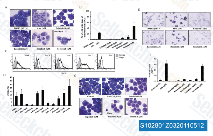
Datos de [ Biochem Pharmacol , 2011 , 82, 1457-1466 ]
-
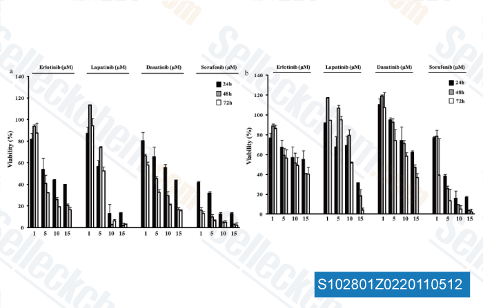
Datos de [ Biochem Pharmacol , 2011 , 82, 1457-1466 ]
-
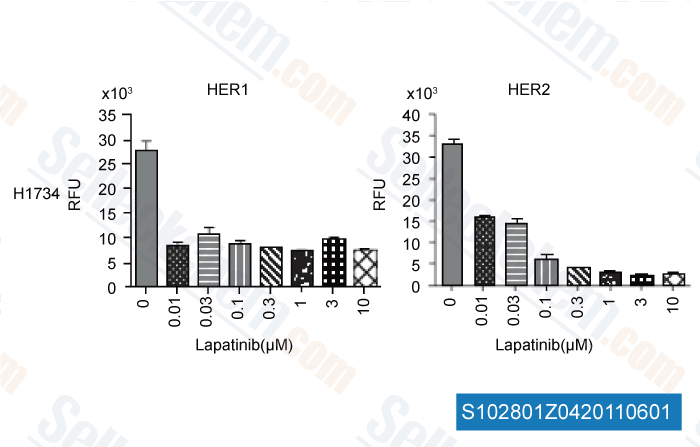
Datos de [ Int J Proteomics , 2011 , 2011, 215496 ]
Sellecks Lapatinib Ditosylate Ha sido citado por 149 Publicaciones
| Inactivation of necroptosis-promoting protein MLKL creates a therapeutic vulnerability in colorectal cancer cells [ Cell Death Dis, 2025, 16(1):118] | PubMed: 39979285 |
| Multiscale Modeling Uncovers Macrophage Infiltration and TNF-α Signaling Networks for Targeting in Inflammatory Breast Cancer Tumor Emboli [ bioRxiv, 2025, 2025.05.29.656249] | PubMed: 40502021 |
| Altered ribosomal profile in acquired resistance and reversal associates with pathological response to chemotherapy in inflammatory breast cancer [ NPJ Breast Cancer, 2024, 10(1):65] | PubMed: 39075068 |
| Patient-derived rhabdomyosarcoma cells recapitulate the genetic and transcriptomic landscapes of primary tumors [ iScience, 2024, 27(10):110862] | PubMed: 39319271 |
| Profiling of ERBB receptors and downstream pathways reveals selectivity and hidden properties of ERBB4 antagonists [ iScience, 2024, 27(2):108839] | PubMed: 38303712 |
| Massively parallel reporter assays identify enhancer elements in oesophageal Adenocarcinoma [ NAR Cancer, 2024, 6(4):zcae041] | PubMed: 39417090 |
| Overcoming brain-derived therapeutic resistance in HER2+ breast cancer brain metastasis [ bioRxiv, 2024, 2024.02.19.581073] | PubMed: 38529509 |
| Proteomic Assessment of SKBR3/HER2+ Breast Cancer Cellular Response to Lapatinib and Investigational Ipatasertib Kinase Inhibitors [ bioRxiv, 2024, 2024.04.02.587656] | PubMed: 38617302 |
| Protocol for identifying properties of ERBB receptor antagonists using the barcoded ERBBprofiler assay [ STAR Protoc, 2024, 5(2):102987] | PubMed: 38635397 |
| Analysis and modeling of cancer drug responses using cell cycle phase-specific rate effects [ Nat Commun, 2023, 14(1):3450] | PubMed: 37301933 |
POLÍTICA DE DEVOLUCIÓN
La Política de Devolución Incondicional de Selleck Chemical garantiza una experiencia de compra en línea fluida para nuestros clientes. Si no está satisfecho con su compra de alguna manera, puede devolver cualquier artículo(s) dentro de los 7 días posteriores a su recepción. En caso de problemas de calidad del producto, ya sean problemas relacionados con el protocolo o con el producto, puede devolver cualquier artículo(s) dentro de los 365 días a partir de la fecha de compra original. Siga las instrucciones a continuación al devolver productos.
ENVÍO Y ALMACENAMIENTO
Los productos Selleck se transportan a temperatura ambiente. Si recibe el producto a temperatura ambiente, tenga la seguridad de que el Departamento de Inspección de Calidad de Selleck ha realizado experimentos para verificar que la colocación a temperatura normal durante un mes no afectará la actividad biológica de los productos en polvo. Después de la recogida, guarde el producto de acuerdo con los requisitos descritos en la hoja de datos. La mayoría de los productos Selleck son estables en las condiciones recomendadas.
NO PARA USO HUMANO, DIAGNÓSTICO VETERINARIO O TERAPÉUTICO.