|
Hoe te citeren 1. Voor in-tekst citatie (materialen en methoden): 2. Voor de tabel met belangrijke bronnen: |
||
|
Gratis nummer: (877) 796-6397 -- Alleen VS en Canada -- |
Fax: +1-832-582-8590 Bestellingen: +1-832-582-8158 |
Technische ondersteuning: +1-832-582-8158 Ext:3 Gelieve uw bestelnummer in de e-mail te vermelden. Wij streven ernaar alle e-mailvragen binnen één werkdag te beantwoorden. |
Biologische Beschrijving
| Specificiteit | MCP1 Antibody [M3B19] detecteert endogene niveaus van totaal MCP1-eiwit. |
|---|---|
| Achtergrond | Monocyt chemoattractant proteïne-1 (MCP-1/CCL2) is een lid van de C-C chemokinfamilie en functioneert als een potente chemotactische factor voor monocyten. Het wordt beschouwd als identiek aan JE, een gen dat oorspronkelijk in muis fibroblasten werd geïdentificeerd als geïnduceerd door bloedplaatjes-afgeleide groeifactor. Het menselijke MCP-1 gen bevindt zich op chromosoom 17q11.2 en codeert voor een eiwit van 76 aminozuren met een molecuulgewicht van ongeveer 13 kDa. MCP-1 behoort tot een subfamilie van chemokines die ten minste vier leden omvat: MCP-1, MCP-2, MCP-3 en MCP-4. CCL2 wordt geproduceerd door een breed scala aan celtypen, hetzij constitutief of als reactie op stimuli zoals oxidatieve stress, cytokines en groeifactoren. De bronnen omvatten endotheelcellen, fibroblasten, epitheelcellen, gladde spiercellen, mesangiale cellen, astrocyten, monocyten en microglia – celtypen die belangrijke rollen spelen in de antivirale immuunafweer in zowel de bloedsomloop als de weefsels. Functioneel gezien stuurt CCL2 de migratie en infiltratie van monocyten, geheugen-T-cellen en natural killer (NK)-cellen. De biologische effecten van CCL2 worden gemedieerd via zijn receptor, CCR2, waarvan de expressie beperkter is in vergelijking met die van CCL2. CCR2 bestaat in twee alternatief gesplicede isovormen, CCR2A en CCR2B, die alleen verschillen in hun C-terminale regio's. |
Gebruiksinformatie
| Toepassing | IHC | Verdunning |
|
|---|---|---|---|
| Reactiviteit | Mouse | ||
| Bron | Rat Monoclonal Antibody | MW | 11 kDa |
| Opslagbuffer | PBS, pH 7.2+50% Glycerol+0.05% BSA+0.01% NaN3 | Opslag (Vanaf de datum van ontvangst) |
-20°C (avoid freeze-thaw cycles), 2 years |
| IHC |
Experimental Protocol:
Deparaffinization/Rehydration
1. Deparaffinize/hydrate sections:
2. Incubate sections in three washes of xylene for 5 min each.
3. Incubate sections in two washes of 100% ethanol for 10 min each.
4. Incubate sections in two washes of 95% ethanol for 10 min each.
5. Wash sections two times in dH2O for 5 min each.
6.Antigen retrieval: For Citrate: Heat slides in a microwave submersed in 1X citrate unmasking solution until boiling is initiated; continue with 10 min at a sub-boiling temperature (95°-98°C). Cool slides on bench top for 30 min.
Staining
1. Wash sections in dH2O three times for 5 min each.
2. Incubate sections in 3% hydrogen peroxide for 10 min.
3. Wash sections in dH2O two times for 5 min each.
4. Wash sections in wash buffer for 5 min.
5. Block each section with 100–400 µl of blocking solution for 1 hr at room temperature.
6. Remove blocking solution and add 100–400 µl primary antibody diluent in to each section. Incubate overnight at 4°C.
7. Remove antibody solution and wash sections with wash buffer three times for 5 min each.
8. Cover section with 1–3 drops HRPas needed. Incubate in a humidified chamber for 30 min at room temperature.
9. Wash sections three times with wash buffer for 5 min each.
10. Add DAB Chromogen Concentrate to DAB Diluent and mix well before use.
11. Apply 100–400 µl DAB to each section and monitor closely. 1–10 min generally provides an acceptable staining intensity.
12. Immerse slides in dH2O.
13. If desired, counterstain sections with hematoxylin.
14. Wash sections in dH2O two times for 5 min each.
15. Dehydrate sections: Incubate sections in 95% ethanol two times for 10 sec each; Repeat in 100% ethanol, incubating sections two times for 10 sec each; Repeat in xylene, incubating sections two times for 10 sec each.
16. Mount sections with coverslips and mounting medium.
|
Referenties
|
Toepassingsgegevens
IHC
Gevalideerd door Selleck
-
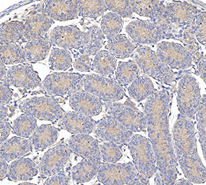 Immunohistochemical analysis of formalin fixed paraffin embedded mouse testicles tissue with F3225 at 1:10 dilution.